


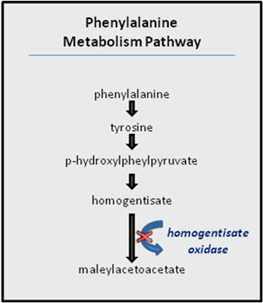
 Figure 1: Phenylalanine metabolism pathway.


« Prev Next »
In 1902, Archibald Garrod published his observations regarding patients whose urine turned black. In patients with this condition, known as alkaptonuria, there is buildup of a chemical called homogentisate, which causes the darkening of the urine. Homogentisate accumulation can lead to extreme joint pain and deterioration so severe that joint replacement is typically needed by the age of 50 in the modern clinical environment. Garrod's knowledge of biochemistry, specifically of the biochemical pathway that produces homogentisate, helped him propose an important hypothesis explaining the underlying cause of the black urine condition.
Normally, excess amounts of the amino acid phenylalanine are metabolized by the human body through a four-step biochemical reaction that yields maleylacetoacetate (Figure 1). The presence of the intermediate metabolite homogentisate in affected patients' urine led Garrod to surmise that the enzyme responsible for its breakdown in the phenylalanine metabolism pathway, homogentisate oxidase, must be defective in these patients. Moreover, because the black urine phenotype followed a Mendelian recessive inheritance pattern, Garrod reasoned that a gene must be responsible for the production of the defective enzyme. Homogentisate's place in the phenylalanine metabolism pathway further suggested that the condition could be linked with other genetic disorders, such as phenylketonuria (PKU) and tyrosinosis.
Garrod's proposition, attributing a defective enzyme to a defective gene, was the first ever to suggest a direct link between genes and proteins. Given that the nucleus was known to house the genetic material, which had already been identified as deoxyribonucleic acid (DNA), several investigators subsequently suggested that the nucleus could also be the central area of protein assembly. A series of seminal experiments, including those summarized in the following sections, disproved this initial supposition about where proteins are made.
The Nucleus and Protein Synthesis
The single-celled alga Acetabularia played an important role in elucidating the site of protein synthesis. During part of its life cycle, Acetabularia takes on an unusual umbrella-like form, during which the cell's nucleus is located completely within the boundaries of a spatially distinct "foot" called a rhizoid. This arrangement serendipitously creates an opportunity in which the nucleus can be removed without causing major damage to the cell. Then, after removal of the nucleus-containing rhizoid, the cell's protein production can be measured over time.
If the nucleus is the site of protein production, then such production should immediately cease in Acetabularia upon the removal of the rhizoid. However, researchers unexpectedly discovered that enucleated Acetabularia cells can survive for months, although protein synthesis in these cells only continues for about two weeks following rhizoid removal (Hämmerling 1953, 1963). Because this experiment demonstrated that a cell's ability to produce new protein does not end as soon as its nucleus is removed, it also demonstrated that the nucleus is not the primary protein-producing cellular organelle. Nonetheless, this study did suggest some other role for the nucleus in the long-term ability to produce proteins.
The Missing Link Between DNA and Proteins

Figure 2: Growing polysomes on a chromosome from E. coli.
The arrow designates where investigators believe RNA polymerase is sitting at, or near, the initiation site for a gene. Along the chromosome, growing polysomes can be seen.
© 1970 American Association for the Advancement of Science. Miller, O. L. et al. Visualization of bacterial genes in action. Science 169, 392–395 (1970). All rights reserved. 
Although Garrod and several other scientists had demonstrated a clear association between genes (which were known to be on chromosomes in the nucleus) and proteins, the precise nature of this link remained mysterious for some time. Researchers wondered whether chromosomes participated directly in protein production. If so, one would expect that some DNA would be found beyond the nucleus, in the cytoplasm, at least some of the time. However, no evidence of DNA outside the nucleus had ever been found. Thus, the exclusive localization of DNA to the nucleus could only be linked to protein synthesis in the cytoplasm if there were some kind of intermediate messenger—a substance "between" the DNA in the nucleus and the protein production machinery in the cytoplasm. The early work of Brachet (1942) and others suggested that another type of nucleic acid, ribonucleic acid (RNA), might be the intermediary. Several pieces of evidence implicated RNA in protein production, including the following:
- RNA is found in both the nucleus and the cytoplasm.
- RNA concentration correlates with protein production.
- Cells that produce large amounts of protein have cytoplasmic dye- and radiation-absorbing regions indicative of the presence of nucleic acids.
- Treatment of cells with ribonuclease (an enzyme that breaks down RNA) decreases the cells' dye- and radiation-absorbing regions.
The identification of RNA as the crucial missing link between DNA and protein was subsequently confirmed by cell fractionation experiments, in which the different parts of cells were separated and examined. In these experiments, the cells were lysed, or opened, with detergents and then spun with high-speed centrifugation, which separated the different cellular components with distinct physical characteristics. In this way, researchers obtained a series of distinct fractions, the heaviest of which included the cells' nuclei, followed by a mitochondrial fraction, then a fraction including other membranous organelles (such as the endoplasmic reticulum), and finally a microsomal fraction of smaller particles. When these different fractions were examined for protein production activity, it was found that most of the new protein synthesis was associated not with the organelle fractions, but rather with the microsomal fraction.
Identifying the source of protein synthesis within the microsomal fraction required an extremely close look at that fraction's components. When the microsomal fraction was examined by electron microscopy both before and after treatment with RNAse, researchers noted that most of the RNA in the fraction was associated with small punctate organelles now known as ribosomes (Figure 2). Ribosomes have since been confirmed to be the "workbenches" for protein synthesis. Later electron microscopy showed that ribosomes are distributed along messenger RNA (mRNA) strands like pearls on a string, thereby enabling multiple copies of a protein to be synthesized simultaneously. The cluster of mRNA-bound ribosomes is called the polyribosome, or polysome for short.
Work by Brenner, Jacob, and Meselson (1961) suggested that the mRNA attached to ribosomes was formed as a sort of disposable copy of the DNA, meant to serve the role of messenger between the DNA in the nucleus and the ribosomes in the cytoplasm. Indeed, mRNA molecules do originate in the nucleus, where they are built based on DNA sequences that serve as templates in their synthesis. The mRNA molecules then diffuse from the nucleus out to the cytoplasm, where they associate with ribosomes and have their genetic messages translated into proteins. This vital series of processes that brings the genetic code from DNA through mRNA to finished proteins has become the central dogma of modern molecular biology.
References and Recommended Reading
Brachet, J. La localisation des acides pentosenucléiques dans les tissus animaux et les oeufs d'Amphibiens en voie de développement. Archives de Biologie 53, 207–257 (1942)
Brenner, S., et al. An unstable intermediate carrying information from genes to ribosomes for protein synthesis. Nature 190, 576–581 (1961)
Garrod, A. E. The incidence of alkaptonuria: A study of chemical individuality. Lancet 2, 1616–1620 (1902).
Hämmerling, J. Nucleo-cytoplasmic relationships in the development of Acetabularia. International Review of Cytology 2, 475–498 (1953)
———. Nucleo-cytoplasmic interactions in Acetabularia and other cells. Annual Review of Plant Physiology 14, 65–92 (1963)
Miller, O. L., et al. Visualization of bacterial genes in action. Science 169, 392–395 (1970)
Prasad, C., & Galbraith, P. A. Sir Archibald Garrod and alkaptonuria: "Story of metabolic genetics." Clinical Genetics 68, 199–203 (2005)
Robinson, A. D. An evaluation of Garrod's contribution to the one gene-one enzyme hypothesis. BioScience 24, 357-358 (1974)
Scriver, C. R. Alkaptonuria:
Such a long journey. Nature Genetics 14, 5-6 (1996) (link to article)


