
Volume 28 Issue 2, February 2023
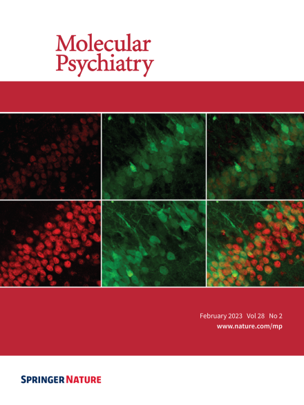
A representative immunofluorescence image showing knockdown of endogenous TDP-43 (upper left, red) from CA1 neurons in adult mouse hippocampus by Adeno-associated virus encoding an shRNA targeting TDP-43. The endogenous levels of TDP-43 were measured from CA1 neurons expressing an shRNA targeting firefly luciferase as a control (lower left, red). GFP was co-expressed with shRNAs and labeled the infected neurons (middle two images, green). Overlaid images of TDP-43 and GFP were shown on the right. For more information see the article by Ni et al. on pages 931-945.
Image
-
Advertisement
