


 Figure 2
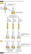


« Prev Next »
In the first half of the twentieth century, Gregor Mendel's principles of genetic inheritance became widely accepted, but the chemical nature of the hereditary material remained unknown. Scientists did know that genes were located on chromosomes and that chromosomes consisted of DNA and proteins. At the time, however, proteins seemed to be a better choice for the genetic material, because chemical analyses had shown that proteins are more varied than DNA in their chemical composition, as well as in their physical properties. Therefore, the eventual identification of DNA as the hereditary material came as a surprise to scientists. This breakthrough resulted from a series of experiments with bacteria and bacteriophages, or viruses that infect bacteria. Together, these experiments demonstrated that DNA was transferred between generations and that this molecule had the ability to transform the properties of a cell.
Frederick Griffith Discovers Bacterial Transformation
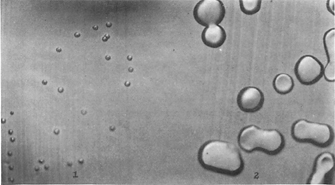
Figure 1: R variant phenotypes.
In this figure the colonies of R variant on the left are grown on agar in the absence of the transforming substance (x 3.5) and on the right are colonies after induction of the transforming substance.
Creative Commons Avery, O. T. et al. Studies on the chemical nature of the substance inducing transformation of pneumococcal types. Journal of Experimental Medicine. 79, 137-157 (1944). 
In the aftermath of the deadly 1918 flu epidemic, governments across the globe rushed to develop vaccines that could stop the spread of infectious diseases. In England, microbiologist Frederick Griffith was studying two strains of Streptococcus pneumoniae that varied dramatically in both their appearance and their virulence, or their ability to cause disease. Specifically, the highly virulent S strain had a smooth capsule, or outer coat composed of polysaccharides, while the nonvirulent R strain had a rough appearance and lacked a capsule (Figure 1). Mice injected with the S strain died within a few days after injection, while mice injected with the R strain did not die.
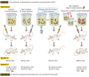
Through a series of experiments, Griffith established that the virulence of the S strain was destroyed by heating the bacteria. Thus, he was surprised to find that mice died when they were injected with a mixture of heat-killed S bacteria and living R bacteria (Figure 2), neither of which caused mice to die when they were injected alone. Griffith was able to isolate live bacteria from the hearts of the dead animals that had been injected with the mixed strains, and he observed that these bacteria had the smooth capsules characteristic of the S strain. Based on these observations, Griffith hypothesized that a chemical component from the virulent S cells had somehow transformed the R cells into the more virulent S form (Griffith, 1928). Unfortunately, Griffith was not able to identify the chemical nature of this "transforming principle" beyond the fact that it was able to survive heat treatment.
Looking back on Griffith's results, which were published in 1928, the interpretation seems obvious. Today, we know that DNA molecules can renature after heat treatment and that bacteria can take up foreign DNA from the environment by a process that we still refer to as transformation. These facts would not be discovered, however, until other scientists conducted further explorations of the nature and function of DNA.
DNA Is Identified as the “Transforming Principle”
The actual identification of DNA as the "transforming principle" was an unexpected outcome of a series of clinical investigations of pneumococcal infections performed over many years (Steinman & Moberg, 1994). At the same time that Griffith was conducting his experiments, researcher Oswald Avery and his colleagues at the Rockefeller University in New York were performing detailed analyses of the pneumococcal cell capsule and the role of this capsule in infections. Modern antibiotics had not yet been discovered, and Avery was convinced that a detailed understanding of the pneumococcal cell was essential to the effective treatment of bacterial pneumonia. Over the years, Avery's group had accumulated considerable biochemical expertise as they established that strains of pneumococci could be distinguished by the polysaccharides in their capsules and that the integrity of the capsule was essential for virulence. Thus, when Griffith's results were published, Avery and his colleagues recognized the importance of these findings, and they decided to use their expertise to identify the specific molecules that could transform a nonencapsulated bacterium into an encapsulated form. In a significant departure from Griffith's procedure, however, Avery's team employed a method for transforming bacteria in cultures rather than in living mice, which gave them better control of their experiments.
Avery and his colleagues, including researchers Colin MacLeod and Maclyn McCarty, used a process of elimination to identify the transforming principle (Avery et al., 1944). In their experiments (Figure 3), identical extracts from heat-treated S cells were first treated with hydrolytic enzymes that specifically destroyed protein, RNA, or DNA. After the enzyme treatments, the treated extracts were then mixed with live R cells. Encapsulated S cells appeared in all of the cultures, except those in which the S strain extract had been treated with DNAse, an enzyme that destroys DNA. These results suggested that DNA was the molecule responsible for transformation.
Avery and his colleagues provided further confirmation for this hypothesis by chemically isolating DNA from the cell extract and showing that it possessed the same transforming ability as the heat-treated extract. We now consider these experiments, which were published in 1944, as providing definitive proof that DNA is the hereditary material. However, the team's results were not well received at the time, most likely because popular opinion still favored protein as the hereditary material.
Hershey and Chase Prove Protein Is Not the Hereditary Material
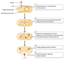
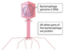
Protein was finally excluded as the hereditary material following a series of experiments published by Alfred Hershey and Martha Chase in 1952. These experiments involved the T2 bacteriophage, a virus that infects the E. coli bacterium. At the time, bacteriophages were widely used as experimental models for studying genetic transmission because they reproduce rapidly and can be easily harvested. In fact, during just one infection cycle, bacteriophages multiply so rapidly within their host bacterial cells that they ultimately cause the cells to burst, thus releasing large numbers of new infectious bacteriophages (Figure 4). The T2 bacteriophage used by Hershey and Chase was known to consist of both protein and DNA, but the role that each substance played in the growth of the bacteriophage was unclear. Electron micrographs had shown that T2 bacteriophages consist of an icosahedral head, a cylindrical sheath, and a base plate that mediates attachment to the bacterium, shown schematically in Figure 5. After infection, phage particles remain attached to the bacterium, but the heads appear empty, forming "ghosts."
To determine the roles that the T2 bacteriophage's DNA and protein play in infection, Hershey and Chase decided to use radioisotopes to trace the fate of the phage's protein and DNA by taking advantage of their chemical differences. Proteins contain sulfur, but DNA does not. Conversely, DNA contains phosphate, but proteins do not. Thus, when infected bacteria are grown in the presence of radioactive forms of phosphate (32P) or sulfur (35S), radioactivity can be selectively incorporated into either DNA or protein. Hershey and Chase employed this method to prepare both 32P-labeled and 35S-labeled bacteriophages, which they then used to infect bacteria. To determine which of the labeled molecules entered the infected bacteria, they detached the phage ghosts from the infected cells by mechanically shearing them off in an ordinary kitchen blender. The ghosts and bacterial cells were then physically separated using a centrifuge. The larger bacterial cells moved rapidly to the bottom of the centrifuge tube, where they formed a pellet. The smaller, lighter phage ghosts remained in the supernatant, where they could be collected and analyzed. During analysis, Hershey and Chase discovered that almost all of the radioactive sulfur remained with the ghosts, while about one-third of the radioactive phosphate entered the bacterial cells and could later be recovered in the next generation of bacteriophages.
From these experiments, Hershey and Chase determined that protein formed a protective coat around the bacteriophage that functioned in both phage attachment to the bacterium and in the injection of phage DNA into the cell. Interestingly, they did not conclude that DNA was the hereditary material, pointing out that further experiments were required to establish the role that DNA played in phage replication. In fact, Hershey and Chase circumspectly ended their paper with the following statement: "This protein probably has no function in the growth of intracellular phage. The DNA has some function. Further chemical inferences should not be drawn from the experiments presented" (Hershey & Chase, 1952). However, a mere one year later, the structure of DNA was determined, and this allowed investigators to put together the pieces in the question of DNA structure and function.
References and Recommended Reading
Avery, O. T., et al. Studies on the chemical nature of the substance inducing transformation of pneumococcal types. Journal of Experimental Medicine 79, 137–157 (1944)
Griffith, F. The significance of pneumococcal types. Journal of Hygiene 27, 113–159 (1928)
Hershey, A. D., & Chase, M. Independent functions of viral protein and nucleic acid in growth of bacteriophage. Journal of General Physiology 36, 39–56 (1952)
Steinman, R. M., & Moberg, C. L. A triple tribute to the experiment that transformed biology. Journal of Experimental Medicine 179, 379–384 (1994)


