Thank you for visiting nature.com. You are using a browser version with limited support for CSS. To obtain
the best experience, we recommend you use a more up to date browser (or turn off compatibility mode in
Internet Explorer). In the meantime, to ensure continued support, we are displaying the site without styles
and JavaScript.
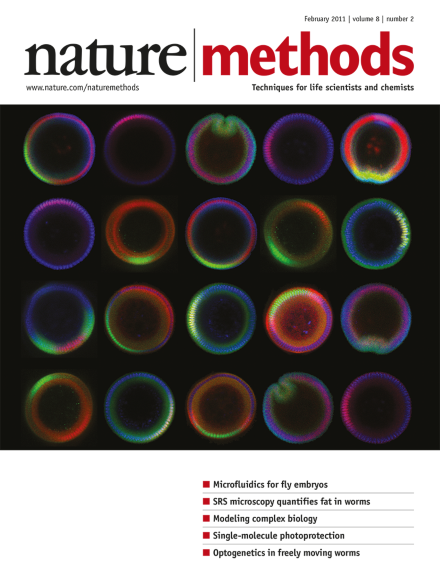
Confocal cross-sections of vertically oriented Drosophila melanogaster embryos in a microfluidic embryo-trap array. Cover design by Erin Dewalt, based on an image provided by Hang Lu. Article p171
In recent years, single-molecule force spectroscopy techniques have been used to study how inter- and intramolecular interactions control the assembly and functional state of biomolecular machinery in vitro. Here we discuss the problems and challenges that need to be addressed to bring these technologies into living cells and to learn how cellular machinery is controlled in vivo.
Combining rule-based descriptions of biochemical reactions with agent-based computer simulation opens new avenues for exploring complex cellular processes.
Stimulated Raman scattering (SRS) microscopy is used to directly visualize lipids in cells and model organisms, and facilitates screening for genes involved in fat storage.
Stimulated Raman scattering (SRS) microscopy is a quantitative, label-free imaging method to map fat distribution and accumulation with high spatial resolution and sensitivity at both cellular and organism levels.
In vivo calcium imaging at multiple depths simultaneously is shown using multifocal two-photon microscopy and spatiotemporal multiplexing. This technique involves scanning the sample with multiple beams in parallel at different axial planes and is applied to monitor neuronal network activity in multiple cortical layers of an anesthetized mouse.
Single-molecule fluorescence experiments with microsecond time resolution are made possible using a photoprotection cocktail that reduces dye blinking and bleaching with a combination of dissolved oxygen, a triplet quencher and a free-radical scavenger.
An optogenetic illumination system based on the use of a digital micromirror device and video tracking software is reported, which allows real-time light delivery with high spatial resolution to specified targets in freely moving Caenorhabditis elegans. Also in this issue, Stirman et al. report a similar illumination system using a liquid crystal display projector. Both methods allow optogenetic perturbation of a variety of neural circuits in the behaving worm.
An optogenetic illumination system based on the use of a liquid crystal display projector and video tracking software is reported, which allows real-time multispectral light delivery with high spatial resolution to specified targets in freely moving Caenorhabditis elegans. Also in this issue, Leifer et al. report a similar illumination system using a digital micromirror device. Both methods allow optogenetic perturbation of a variety of neural circuits in the behaving worm.
By individually replacing 16 yeast genes encoding ABC transporters by GFP, mating and selecting for strains with accumulated mutations the authors create a Green Monster, a strain with deletions in all 16 genes.
A microfluidic embryo-trap array for large-scale end-on imaging of Drosophila melanogaster embryos permits quantitative analysis of dorsoventral gradients during development.
The Network-Free Stochastic Simulator (NFsim) allows the representation of complex biological systems as rule-based models and facilitates coarse-graining of the reaction mechanisms.