Volume 8
-
No. 12 December 2011
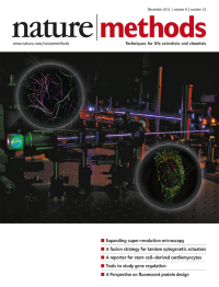
A photograph of the optical setup that combines photoactivated localization microscopy and light-sheet microscopy. Floating above this are fluorescence images obtained using two other advanced imaging techniques, stochastic optical reconstruction microscopy and structured illumination microscopy. Analysis p1027, Brief Communications p1044, p1047
-
No. 11 November 2011
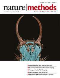
This photograph of a green lacewing larva (Chrysopa sp.) took first place in the 2011 Nikon Small World Photomicrography Competition. The image was taken by Igor Siwanowicz of the Max Planck Institute of Neurobiology, using confocal microscopy at 20× magnification. The full gallery of winning images can be viewed at http://www.nikonsmallworld.com/.
-
No. 10 October 2011
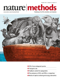
The cover image depicts in abstract fashion the preservation of the genomes of animal species using technology. Although it refers to a paper published in this issue (Ben-Nun et al.), it does not intend to suggest that the methods described in themselves permit species preservation. Cover design by Erin Dewalt
-
No. 9 September 2011
The cover image abstractly depicts that zinc-finger nucleases cleave not only the site in the genome they are designed to target but also cleave off-target sites. Off-target cleavage is, however, much less efficient. Cover design by Erin Dewalt. Article p765
-
No. 8 August 2011
We present a special feature in this issue to celebrate 2011 as the International Year of Chemistry. The most important elements for life are indicated in gold on this periodic table (source, the 21 January 2011 version of the International Union of Pure and Applied Chemistry Periodic Table of the Elements). Cover by Erin Dewalt. Special feature p633, Editorial p607, Technology Feature p623
-
No. 7 July 2011
Artistic rendering of the three-dimensional crystal structure of an IgG2A antibody molecule (Protein Data Bank code 11GT). The picture was created with PyMOL and Adobe Photoshop programs by Frederic A. Fellouse. Cover by Erin Dewalt. Analysis p551
-
No. 6 June 2011
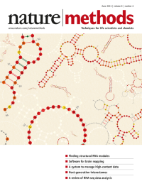
Examples of three-dimensional structural RNA modules predicted by the program RMDetect. Artistic interpretation by Erin Dewalt, based on figures from José Almeida Cruz and Eric Westhof. Article p513
-
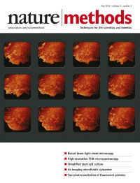
No. 5 May 2011
Three-dimensional time-lapse images of membrane ruffles in COS-7 cells transfected with plasmids encoding mEmerald–c-Src obtained by Bessel beam two-photon light-sheet microscopy. Cover design by Erin Dewalt, based on images provided by James A. Galbraith. Article p417
-
No. 4 April 2011
The cover image artistically depicts the isolation and analysis of a single cell from within a heterogeneous cellular population. Cover design by Erin Dewalt. Supplement Foreword p307
-
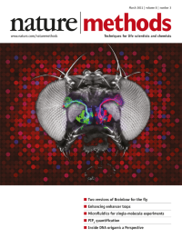
No. 3 March 2011
A depiction of multicolor labeling of projection neurons in the fly olfactory system with Drosophila Brainbow or dBrainbow. A partial projection of a confocal image stack corresponding to three neuronal lineages labeled with fluorescent antibodies is shown superimposed on a bright-field image of a fly head, in homage to an image in a review by Martin Heisenberg (Nat. Rev. Neurosci. 4, 266–275; 2003). Cover by Erin Dewalt based on a figure provided by Stefanie Hampel, Phuong Chung, Andrew M. Seeds and Julie H. Simpson. Article p253
-
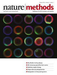
No. 2 February 2011
Confocal cross-sections of vertically oriented Drosophila melanogaster embryos in a microfluidic embryo-trap array. Cover design by Erin Dewalt, based on an image provided by Hang Lu. Article p171
-
No. 1 January 2011
Optogenetics, our pick for Method of the Year 2010, is rapidly becoming a vital tool for neurobiologists, who are applying it to study both basic biology and disease. Cover design by Erin Dewalt. Special feature starts on p19.
Special