Thank you for visiting nature.com. You are using a browser version with limited support for CSS. To obtain
the best experience, we recommend you use a more up to date browser (or turn off compatibility mode in
Internet Explorer). In the meantime, to ensure continued support, we are displaying the site without styles
and JavaScript.
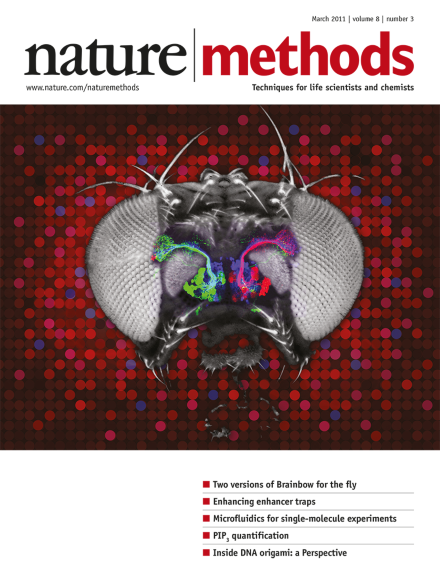
A depiction of multicolor labeling of projection neurons in the fly olfactory system with Drosophila Brainbow or dBrainbow. A partial projection of a confocal image stack corresponding to three neuronal lineages labeled with fluorescent antibodies is shown superimposed on a bright-field image of a fly head, in homage to an image in a review by Martin Heisenberg (Nat. Rev. Neurosci. 4, 266–275; 2003). Cover by Erin Dewalt based on a figure provided by Stefanie Hampel, Phuong Chung, Andrew M. Seeds and Julie H. Simpson. Article p253
An optimized transcription activator–like effector (TALE) and an improved assembly method promise efficient genome editing and transcriptome modulation.
The comprehensive mapping of transcripts, histone modifications and transcription factor binding allows for the functional annotation of fly and worm genomes.
Three single-molecule methods promise to increase the time resolution of experiments, to allow better access to sparsely populated molecular states and to permit combinatorial high-throughput analysis.
Brainbow is a powerful genetic tool for multicolor labeling in mice with applications in fields including developmental biology and neuroanatomy. Now two groups have ported the approach to the fruit fly where it may have even greater impact.
A genetic platform allows refinement of tissue-specific expression using the upstream activating sequence–GAL4 system in Drosophila melanogaster, facilitating the segmentation of complex expression patterns and allowing GAL4 expression patterns to be repurposed.
A laminar flow mixing microfluidic device enables single-molecule fluorescence resonance energy transfer (FRET) kinetic measurements with a time resolution of 0.2 ms, enabling the study of early binding-coupled folding and unfolding events of an intrinsically disordered protein, α-synuclein. Also in this issue, Kim et al. describe another microfluidic mixing device for single-molecule experiments.
A microfluidic mixing device for multiple, rapid and automated single-molecule measurements permits the study of macromolecule properties under varying environmental conditions. Also in this issue, Gambin et al. present another microfluidic mixing device for rapid single-molecule measurements.
Microscope control software allowed automatic machine learning-based detection of rare events in living cells and unattended operation of complex imaging assays. Performance was demonstrated by detailed analysis of processes during transient mitotic stages.
Changing the codon sequence in Caenorhabditis elegans genes allows fine-tuning of transgene expression from high to low expression. The same strategy is likely applicable for Drosophila melanogaster and Saccharomyces cerevisiae.
A genetic multicolor cell-labeling technique for Droshophila melanogaster, Drosophila Brainbow, is described and applied to the study of neural circuits. This method implements a variant of the mouse Brainbow strategy in combination with specific neuronal targeting using the Gal-4–upstream activating sequence system to select for epitope-tagged proteins detectable with immunofluorescence. Also in this issue, Hadjieconomou et al. develop a similar strategy, Flybow, to select for membrane-tethered fluorescent proteins.
A genetic multicolor cell-labeling technique for Droshophila melanogaster, Flybow, is described and applied to the study of neural circuits. This method implements a variant of the mouse Brainbow strategy in combination with specific neuronal targeting using the Gal-4–upstream activating sequence system to select for membrane-tethered fluorescent proteins. Also in this issue, Hampel et al. report a similar strategy, Drosophila Brainbow, to select for epitope-tagged proteins detectable via immunofluorescence.
By methylating the phosphate groups of PtdIns(3,4,5)P3 researchers can load this lipid more efficiently into a mass spectrometer and thus this lipid can be quantified in the presence of an internal synthetic standard.