


 Figure 1


« Prev Next »
With the discovery of the molecular structure of the DNA double helix in 1953, researchers turned to the structure of ribonucleic acid (RNA) as the next critical puzzle to be solved on the road to understanding the molecular basis of life. Indeed, RNA may be the only molecule to have inspired the formation of a club, known as the RNA Tie Club, whose members included Nobel Laureates James Watson and Francis Crick, the discoverers of DNA structure, as well as Sydney Brenner, who was awarded the Nobel Prize in 2002 for his work involving gene regulation in the model organism Caenorhabditis elegans. The members of this club, each nicknamed for a particular amino acid, exchanged letters in which they presented various unpublished ideas in an attempt to understand the structure of RNA and how this molecule participates in the building of proteins. During the following 50 years, many questions were answered and many surprises were uncovered.
Early Discoveries of RNA Structure
Today, researchers know that cells contain a variety of forms of RNA—including messenger RNA (mRNA), transfer RNA (tRNA), and ribosomal RNA (rRNA)—and each form is involved in different functions and activities. Messenger RNA is essentially a copy of a section of DNA and serves as a template for the manufacture of one or more proteins. Transfer RNA binds to both mRNA and amino acids (the building blocks of proteins) and brings the correct amino acids into the growing polypeptide chain during protein formation, based on the nucleotide sequence of the mRNA. The process by which proteins are built is called translation. Translation occurs on ribosomes, which are cellular organelles composed of protein and rRNA.
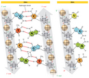
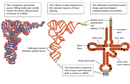
Although there are multiple types of RNA molecules, the basic structure of all RNA is similar. Each kind of RNA is a polymeric molecule made by stringing together individual ribonucleotides, always by adding the 5'-phosphate group of one nucleotide onto the 3'-hydroxyl group of the previous nucleotide. Like DNA, each RNA strand has the same basic structure, composed of nitrogenous bases covalently bound to a sugar-phosphate backbone (Figure 1). However, unlike DNA, RNA is usually a single-stranded molecule. Also, the sugar in RNA is ribose instead of deoxyribose (ribose contains one more hydroxyl group on the second carbon), which accounts for the molecule's name. RNA consists of four nitrogenous bases: adenine, cytosine, uracil, and guanine. Uracil is a pyrimidine that is structurally similar to the thymine, another pyrimidine that is found in DNA. Like thymine, uracil can base-pair with adenine (Figure 2).
Although RNA is a single-stranded molecule, researchers soon discovered that it can form double-stranded structures, which are important to its function. In 1956, Alexander Rich—an X-ray crystallographer and member of the RNA Tie Club—and David Davies, both working at the National Institutes of Health, discovered that single strands of RNA can "hybridize," sticking together to form a double-stranded molecule (Rich & Davies, 1956). Later, in 1960, the discovery that an RNA molecule and a DNA molecule could form a hybrid double helix was the first experimental demonstration of a way in which information could be transferred from DNA to RNA (Rich, 1960).
Single-stranded RNA can also form many secondary structures in which a single RNA molecule folds over and forms hairpin loops, stabilized by intramolecular hydrogen bonds between complementary bases. Such base-pairing of RNA is critical for many RNA functions, such as the ability of tRNA to bind to the correct sequence of mRNA during translation (Figure 3).
Robert Holley, a chemist at Cornell University, was the first researcher to work out the structure of tRNA (Holley et al., 1965). This molecule turned out to be the elusive structure that Francis Crick proposed in his so-called "adapter hypothesis" of 1955—a structure that carried amino acids and arranged them in a certain order that corresponded to the sequence in the nucleic acid strand. In 1968, Holley was awarded the Nobel Prize in Physiology or Medicine together with Gobind Khorana, at the University of Wisconsin, and Marshall Nirenberg, at the National Institutes of Health. Nirenberg and Khorana devised the key experiments to decipher the genetic code—in other words, which sequences of three nucleotides (codons) in an mRNA molecule would code for which amino acids.
mRNA and Splicing
Several forms of RNA play pivotal roles in gene expression—the
process responsible for manifesting the instructions stored in the sequence of
DNA nucleotides in either RNA or protein molecules that carry out the cell's
activities (Figures 4 & 5).
Messenger RNA (mRNA) is particularly important in this process. mRNA is
primarily composed of coding sequences; that is, it carries the genetic
information for the amino acid sequence of a protein to the ribosome, where
that particular protein is synthesized. In addition, each mRNA molecule also
contains noncoding, or untranslated, sequences that may carry instructions for
how the mRNA is handled by the cell (Figure 6).
For example, the untranslated region at the 5' end of the mRNA molecules found
in bacteria and other prokaryotes contains what is called a Shine-Dalgarno
sequence, which aids in the binding of the mRNA to ribosomes.
In contrast, the mRNA of eukaryotic organisms is prepared for translation through more complex mechanisms. For one, the addition of a guanine nucleotide with a methyl (CH3) group to the 5' end of the mRNA, called the 5' cap, increases the stability of the mRNA and assists in the binding of the mRNA to the ribosome for translation. Meanwhile, another untranslated region is added to the 3' end of the mRNA, thereby further affecting the stability of the molecule. In this case, a "tail" consisting of anywhere from 50 to 250 adenine nucleotides is added to the 3' end. This poly(A) tail can increase the stability of many mRNA molecules, depending on the proteins that attach to it. The greater the stability, and the longer an mRNA molecule exists in a cell, the more protein that can be made from that molecule.
In eukaryotes (and to a lesser extent, prokaryotes), when RNA is first transcribed from DNA, it may contain additional noncoding sequences that are interspersed within the coding sequence. This immature RNA molecule is referred to as precursor mRNA (pre-mRNA) or heterogeneous nuclear RNA (hnRNA). The intervening noncoding sequences are called introns, and the segments of coding are known as material exons. The introns are then removed by a process known as RNA splicing to produce the mature mRNA molecule (Figure 7). An organelle called the spliceosome, composed of protein and small nuclear RNAs (snRNAs), is responsible for recognizing and removing the introns from pre-mRNA.
The surprising discovery of RNA splicing caused a paradigm shift in genetics. Much early work indicated that mRNA and the genes in DNA were colinear; that is, they were thought to match up, base for base, with the exception of the 3' poly(A) tail. In the late 1970s, however, seminal studies of gene expression in cells infected with an adenovirus demonstrated that the RNA transcripts produced by viral infection contained sequences that were not next to one another in the viral genome. Further study revealed that these mRNAs were produced after material had been removed or spliced out of a larger primary transcript (Berget et al., 1977; Evans et al., 1977). Since that time, introns have been found to occur in many eukaryotic cellular genes and some prokaryotic genes.
Probably the most thoroughly studied class of introns consists of those found in protein-coding genes. The 5' end of these introns almost always begins with the dinucleotide GU, and the 3' end typically contains AG. Changing one of these nucleotides precludes splicing. Another important sequence occurs at the branch point, anywhere from 18 to 40 nucleotides upstream from the 3' end of an intron. This sequence always contains an adenine, but it is otherwise loosely conserved. A typical sequence at a branch point is YNYYRAY, where Y indicates a pyrimidine, N denotes any nucleotide, R any purine, and A is for adenine (Figure 8) (Pierce, 2000; Patel & Steitz, 2003).
Many eukaryotic genes can be spliced in a number of different ways by choosing between different potential 5′ and 3′ splice junctions, thereby creating different combinations of exons and introns in the final mRNAs. This mix-and-match process allows the creation of several different proteins from a single gene sequence. The first example of such "alternative splicing" (Figure 9) was discovered in the adenovirus in 1977 (Berget et al., 1977). The first example in cellular genes was reported in 1980 in the IgM gene, which encodes an immunoglobulin, one of several proteins created by immune cells to fight infection by foreign organisms and particles (Early et al., 1980).
The Dscam gene of Drosophila, which encodes proteins involved in guiding embryonic nerves to their target destinations during formation of the fly's nervous system, exhibits an especially impressive number of alternative splicing patterns. Dozens of different forms of Dscam mRNAs and corresponding proteins have been identified, while analysis of the gene's sequence reveals a staggering 38,000 potential additional mRNAs, based on the large number of introns found. The ability to produce so many different proteins from a single gene may be necessary for forming as complex a structure as the nervous system (Schmucker et al., 2000). In general, the existence of multiple mRNA transcripts from single genes may account for the complexity of some organisms, such as humans, even though these organisms have relatively few genes (in the case of humans, approximately 25,000).
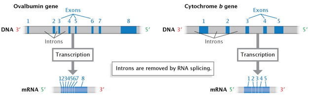
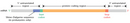
Figure 7: Introns are removed during RNA splicing.
Non-coding sequences, or introns, are removed during RNA splicing to produce a mature mRNA transcript composed of exons (coding sequences).
© 2014 Nature Education Adapted from Pierce, Benjamin. Genetics: A Conceptual Approach, 2nd ed. All rights reserved. 
tRNA and rRNA: Their Role in Translation
Two additional categories of RNA play a critical role in the
translation process: tRNA and rRNA. Ribosomal RNA (rRNA) molecules were
initially characterized by how rapidly they would "sink" in a centrifuge tube—in
other words, they were described by their sedimentation velocity as measured in
Svedberg (S) units. Prokaryotic organisms contain one type of rRNA gene that
encodes three distinct RNA species: the 23S, 5S, and 16S rRNAs. In comparison,
eukaryotic cells contain two types of rRNA genes that give rise to four rRNA
species: the 28S, 5.8S, 5S, and 18S rRNAs. Both the eukaryotic and prokaryotic
genomes contain multiple copies of these rRNA genes to be able to manufacture
the large number of ribosomes required by a cell. Mature rRNAs are produced by
cleavage and modification of initial transcripts (Pierce, 2000).
Transfer RNA (tRNA) molecules serve as molecular adaptors that bind to mRNA on one end and carry amino acids into position on the other. Most types of cells possess approximately 30 to 40 different tRNAs, with more than one tRNA corresponding to each amino acid. tRNAs fold into a cloverleaf structure held together by the pairing of complementary nucleotides. Structural studies using X-ray crystallography have demonstrated that the cloverleaf is further folded into an L shape (Figure 10). A loop at one end of the folded structure base-pairs with three nucleotides on the mRNA that are collectively called a codon; the complementary three nucleotides on the tRNA are called the anticodon.
Although the pairing between codon and anticodon takes place over three nucleotides, strict complementary base-pairing is only necessary between the first two nucleotides. The third position is referred to as the "wobble" position (Figure 11), and the rules for base-pairing are less stringent at this position. Because of this flexibility, the 30 to 40 tRNAs present in a cell can "read" all 61 codons in mRNA.
The opposite end of the folded structure, which is the 3' end of the tRNA, binds to its corresponding amino acid at an attachment site that is also three nucleotides long, invariably CCA. Enzymes called aminoacyl-tRNA synthetases attach the correct amino acid to each tRNA, based on the three-dimensional structure of the tRNA molecule.
More and More RNAs
Finally, there are still more forms of RNA beyond mRNA, rRNA, and tRNA. For instance, short RNAs are not only part of organelles like ribosomes and spliceosomes, but also of some enzymes. For example, the enzyme telomerase, which adds nucleotides to the ends of chromosomes, is composed of a 451-nucleotide RNA and several proteins. Juli Feigon at the University of California, Los Angeles, together with postdoctoral scholar Carla Theimer and graduate student Craig Blois, first solved the structure of an essential piece of this RNA by nuclear magnetic resonance spectroscopy (Theimer et al., 2005). They revealed a unique RNA structure with extensive RNA folding, which is necessary for telomerase activity.
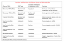
Other classes of RNA species include microRNAs, small interfering RNAs, and sRNAs—all of which are not translated into proteins but still perform important functions in the cell. The discovery of these RNAs has been one of the most exciting advances in recent years, and there is currently a lot of interest in the use of these molecules as possible therapies. But as far as their structure is concerned, these RNAs all share the same basic single-stranded chemical structure with, in some cases, higher-order structures obtained through complementary base-pair folding.
From the RNA Tie Club to today, the more scientists have studied RNA, the more surprises they have uncovered. New functions for RNA, new modifications to RNA, and other surprises undoubtedly await discovery in the years to come.
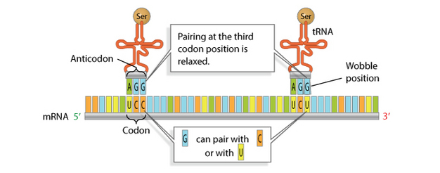
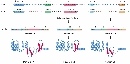
Figure 11: The "wobble" position.
Base-pairing rules between the tRNA anticodon and the mRNA codon are less stringent at the third nucleotide position. This base-pairing flexibility is also called "wobble."
© 2014 Nature Education Adapted from Pierce, Benjamin. Genetics: A Conceptual Approach, 2nd ed. All rights reserved.
References and Recommended Reading
Berget, S. M., Moore, C., & Sharp, P. A. Spliced segments at the 5' terminus of adenovirus 2 late mRNA. Proceedings of the National Academy of Sciences 74, 3171–3175 (1977)
Early, P., et al. Two mRNAs can be produced from a single immunoglobulin u chain by alternative RNS processing pathways. Cell 20, 313–319 (1980)
Evans, R. M., et al. The initiation sites for RNA transcription in Ad2 DNA. Cell 12, 733–739 (1977)
Holley, R. W., et al. Structure of a ribonucleic acid. Science 147, 1462–1465 (1965) doi:10.1126/science.147.3664.1462
Patel,
A. A., & Steitz, J. A. Splicing double: Insights from the second spliceosome.
Nature 4, 960–970 (2003) doi:10.1038/nrm1259 (link to article)
Pierce, B. A. Genetics: A Conceptual Approach, 2nd ed. (New York, Freeman, 2000)
Rich, A. A hybrid helix containing both deoxyribose and ribose polynucleotides and its relation to the transfer of information between the nucleic acids. Proceedings of the National Academy of Sciences 46, 1044–1053 (1960)
Rich, A., & Davies, D. R. A new two-stranded helical structure: Polyadenylic acid and polyuridylic acid.
Journal of the American Chemical Society
78, 3548–3549 (1956) (link to article)
Schmucker, D., et al. Drosophila Dscam is an axon guidance receptor exhibiting extraordinary molecular diversity. Cell 101, 671–684 (2000)
Theimer, C. A., Blois, C. A., & Feigon, J. Structure of the human telomerase RNA pseudoknot reveals conserved tertiary interactions essential for function. Molecular Cell 17, 671–682 (2005)


