Thank you for visiting nature.com. You are using a browser version with limited support for CSS. To obtain
the best experience, we recommend you use a more up to date browser (or turn off compatibility mode in
Internet Explorer). In the meantime, to ensure continued support, we are displaying the site without styles
and JavaScript.
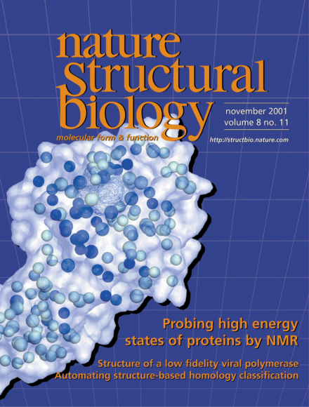
Ligand access to a binding site buried within a protein requires rearrangement of the polypeptide chain; this process likely involves transitions into high energy states. For a mutant T4 lysozyme (surface representation), NMR relaxation experiments reveal that, while most residues in the protein display little motion (white and light blue spheres), the residues lining the ligand-binding cavity (wire frame) undergo significant structural movement (dark blue spheres). See pages 932–935, and News and Views pages 910–912.
A powerful new NMR technique applied to the ubiquitous Ca2+ sensor, calmodulin, reveals significant conformational flexibility within each globular domain, which contributes to its ability to bind a wide range of targets. These measurements of residual dipolar couplings between nuclear spins demonstrate a fast and accurate method for pinpointing structural features that cannot be delineated reliably by traditional NOE analysis.
Many proteins frequently undergo structural rearrangement to complete their functions. Ligand entry and binding are often associated with some degree of localized disorder. Indeed, low populations of disordered excited states may help drive such processes. Characterization of these states is vital to understanding the mechanisms of many biological functions.
Structural characterization of a variety of DNA polymerases has likened the polymerase domain to a hand that grasps DNA with functional subdomains referred to as fingers, palm and thumb. The solution structure of the African swine fever virus DNA polymerase X indicates that it does not have a hand-like architecture and suggests a mechanism by which the polymerase may compensate for the lack of a dedicated DNA binding subdomain.
In the absence of other biological information, the detection of remote homology is a prerequisite step toward understanding the function of a new protein. A novel method based on structure comparison improves our ability to do this automatically and systematically.
The 4.5 Å map of the MsbA protein, a putative lipid A transporter from Escherichia coli, provides the first detailed structural model for the transmembrane domain and cytoplasmic 'loops' of an ABC transporter and the geometric relationship of these regions to the ATP-binding cassette motor domain. Based on this structure, specific hypotheses for the mechanics of the pump can now be formulated and tested.