Thank you for visiting nature.com. You are using a browser version with limited support for CSS. To obtain
the best experience, we recommend you use a more up to date browser (or turn off compatibility mode in
Internet Explorer). In the meantime, to ensure continued support, we are displaying the site without styles
and JavaScript.
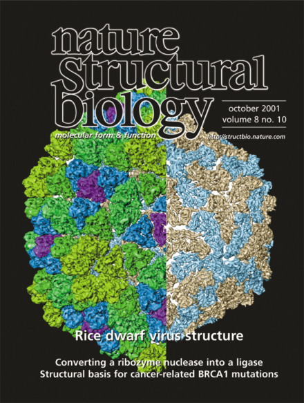
Structure of the rice dwarf virus (RDV) reconstructed from cryo-electron microscopic images. The RDV capsid contains two icosahedral protein shells. The outer shell consists of P8 trimers (colored triangles on the left), whereas the inner shell consists of P3 dimers (each containing one light brown and one light blue subunit in the cutaway view on the right). The 6.8 å resolution map clearly shows the different packing symmetries and structural features ofthe protein subunits in the two shells. See pages 868–873
Death-associated protein kinase (DAP-kinase; DAPk) has been implicated in programmed cell death and tumor suppression. The recently solved crystal structure of the catalytic domain of human DAP-kinase reveals interesting 'fingerprint' regions that may be functionally important.
Crystal structures of the Escherichia coli DNA replication γ clamp loading complex and of a subunit of the clamp loader bound to a β clamp monomer provide a physical framework in which to view ATP-dependent modulation of γ complex–β interactions. The structural data suggest how the β ring is opened and loaded onto DNA in the absence of a direct interaction between the γ complex and the β dimer interface.