Volume 13
-
No. 12 December 2017
Cover image supplied by Dr Farasat Zaman and Prof. Lars Savendahl from the Karolinska Institutet, Stockholm, Sweden. The image shows a complete human epiphyseal (growth) plate extracted from a child after epiphysiodesis of the distal femur. All zones of the growth plate are depicted, including resting cartilage (top) and zones of proliferative and hypertrophic chondrocytes, below which are shown calcified matrix and ossified bone. The tissue was fixed and stained with van Gieson/alcian blue. The extracted growth plate can be used as an ex vivo model to address pre-clinical and clinical questions about human bone development.
-
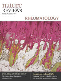
No. 11 November 2017
Cover image supplied by Dr Farasat Zaman and Prof. Lars Savendahl from the Karolinska Institutet, Stockholm, Sweden. The image shows a complete human epiphyseal (growth) plate extracted from a child after epiphysiodesis of the distal femur. All zones of the growth plate are depicted, including resting cartilage (top) and zones of proliferative and hypertrophic chondrocytes, below which are shown calcified matrix and ossified bone. The tissue was fixed and stained with van Gieson/alcian blue. The extracted growth plate can be used as an ex vivo model to address pre-clinical and clinical questions about human bone development.
-
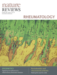
No. 10 October 2017
Cover image supplied by Dr Farasat Zaman and Prof. Lars Savendahl from the Karolinska Institutet, Stockholm, Sweden. The image shows a complete human epiphyseal (growth) plate extracted from a child after epiphysiodesis of the distal femur. All zones of the growth plate are depicted, including resting cartilage (top) and zones of proliferative and hypertrophic chondrocytes, below which are shown calcified matrix and ossified bone. The tissue was fixed and stained with van Gieson/alcian blue. The extracted growth plate can be used as an ex vivo model to address pre-clinical and clinical questions about human bone development.
-
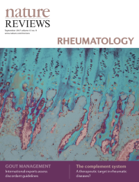
No. 9 September 2017
Cover image supplied by Dr Farasat Zaman and Prof. Lars Savendahl from the Karolinska Institutet, Stockholm, Sweden. The image shows a complete human epiphyseal (growth) plate extracted from a child after epiphysiodesis of the distal femur. All zones of the growth plate are depicted, including resting cartilage (top) and zones of proliferative and hypertrophic chondrocytes, below which are shown calcified matrix and ossified bone. The tissue was fixed and stained with van Gieson/alcian blue. The extracted growth plate can be used as an ex vivo model to address pre-clinical and clinical questions about human bone development.
-
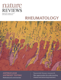
No. 8 August 2017
Cover image supplied by Dr Farasat Zaman and Prof. Lars Savendahl from the Karolinska Institutet, Stockholm, Sweden. The image shows a complete human epiphyseal (growth) plate extracted from a child after epiphysiodesis of the distal femur. All zones of the growth plate are depicted, including resting cartilage (top) and zones of proliferative and hypertrophic chondrocytes, below which are shown calcified matrix and ossified bone. The tissue was fixed and stained with van Gieson/alcian blue. The extracted growth plate can be used as an ex vivo model to address pre-clinical and clinical questions about human bone development.
-
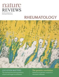
No. 7 July 2017
Cover image supplied by Dr Farasat Zaman and Prof. Lars Savendahl from the Karolinska Institutet, Stockholm, Sweden. The image shows a complete human epiphyseal (growth) plate extracted from a child after epiphysiodesis of the distal femur. All zones of the growth plate are depicted, including resting cartilage (top) and zones of proliferative and hypertrophic chondrocytes, below which are shown calcified matrix and ossified bone. The tissue was fixed and stained with van Gieson/alcian blue. The extracted growth plate can be used as an ex vivo model to address pre-clinical and clinical questions about human bone development.
-
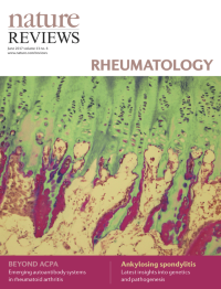
No. 6 June 2017
Cover image supplied by Dr Farasat Zaman and Prof. Lars Savendahl from the Karolinska Institutet, Stockholm, Sweden. The image shows a complete human epiphyseal (growth) plate extracted from a child after epiphysiodesis of the distal femur. All zones of the growth plate are depicted, including resting cartilage (top) and zones of proliferative and hypertrophic chondrocytes, below which are shown calcified matrix and ossified bone. The tissue was fixed and stained with van Gieson/alcian blue. The extracted growth plate can be used as an ex vivo model to address pre-clinical and clinical questions about human bone development.
-
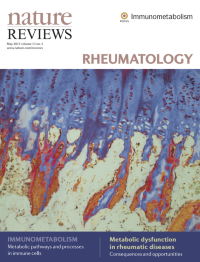
No. 5 May 2017
Cover image supplied by Dr Farasat Zaman and Prof. Lars Savendahl from the Karolinska Institutet, Stockholm, Sweden. The image shows a complete human epiphyseal (growth) plate extracted from a child after epiphysiodesis of the distal femur. All zones of the growth plate are depicted, including resting cartilage (top) and zones of proliferative and hypertrophic chondrocytes, below which are shown calcified matrix and ossified bone. The tissue was fixed and stained with van Gieson/alcian blue. The extracted growth plate can be used as an ex vivo model to address pre-clinical and clinical questions about human bone development.
-
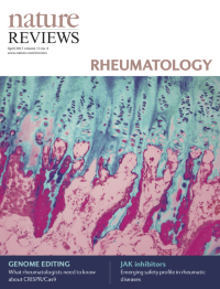
No. 4 April 2017
Cover image supplied by Dr Farasat Zaman and Prof. Lars Savendahl from the Karolinska Institutet, Stockholm, Sweden. The image shows a complete human epiphyseal (growth) plate extracted from a child after epiphysiodesis of the distal femur. All zones of the growth plate are depicted, including resting cartilage (top) and zones of proliferative and hypertrophic chondrocytes, below which are shown calcified matrix and ossified bone. The tissue was fixed and stained with van Gieson/alcian blue. The extracted growth plate can be used as an ex vivo model to address pre-clinical and clinical questions about human bone development.
-
No. 3 March 2017
Cover image supplied by Dr Farasat Zaman and Prof. Lars Savendahl from the Karolinska Institutet, Stockholm, Sweden. The image shows a complete human epiphyseal (growth) plate extracted from a child after epiphysiodesis of the distal femur. All zones of the growth plate are depicted, including resting cartilage (top) and zones of proliferative and hypertrophic chondrocytes, below which are shown calcified matrix and ossified bone. The tissue was fixed and stained with van Gieson/alcian blue. The extracted growth plate can be used as an ex vivo model to address pre-clinical and clinical questions about human bone development.
-
No. 2 February 2017
Cover image supplied by Dr Farasat Zaman and Prof. Lars Savendahl from the Karolinska Institutet, Stockholm, Sweden. The image shows a complete human epiphyseal (growth) plate extracted from a child after epiphysiodesis of the distal femur. All zones of the growth plate are depicted, including resting cartilage (top) and zones of proliferative and hypertrophic chondrocytes, below which are shown calcified matrix and ossified bone. The tissue was fixed and stained with van Gieson/alcian blue. The extracted growth plate can be used as an ex vivo model to address pre-clinical and clinical questions about human bone development.
-
No. 1 January 2017
Cover image supplied by Dr Farasat Zaman and Prof. Lars Savendahl from the Karolinska Institutet, Stockholm, Sweden. The image shows a complete human epiphyseal (growth) plate extracted from a child after epiphysiodesis of the distal femur. All zones of the growth plate are depicted, including resting cartilage (top) and zones of proliferative and hypertrophic chondrocytes, below which are shown calcified matrix and ossified bone. The tissue was fixed and stained with van Gieson/alcian blue. The extracted growth plate can be used as an ex vivo model to address pre-clinical and clinical questions about human bone development.