Thank you for visiting nature.com. You are using a browser version with limited support for CSS. To obtain
the best experience, we recommend you use a more up to date browser (or turn off compatibility mode in
Internet Explorer). In the meantime, to ensure continued support, we are displaying the site without styles
and JavaScript.
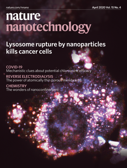
Nanoparticles are usually endocytosed by cells and digested by lysosomes. Therefore, these waste disposal organelles, which are characterized by a low pH as compared to the cytoplasm, offer a potential target for nanoparticle-mediated cancer cell killing. Borkowska et al. designed mixed-charge nanoparticles that, upon entering the acidic tumour microenvironment, aggregate into clusters on the surface of the cells. The nanoparticle clusters are then endocytosed and form even larger clusters in the more acidic lysosomes, as shown in the cover image. Nanoparticle aggregation then causes lysosome swelling and rupture and ultimately leads to cell death. Importantly, the distinct charge decoration of the nanoparticles prevents aggregation in the less acidic extracellular environment of healthy tissues and thus offers a cancer-cell selective targeting strategy.
The identification and isolation of individuals with COVID-19 can help to flatten the epidemic curve and win us time to wait for the vaccine development and production, and antiviral drug therapies.
Chloroquine — an approved malaria drug — is known in nanomedicine research for the investigation of nanoparticle uptake in cells, and may have potential for the treatment of COVID-19.
Although we seem to understand how nanoscience can impact the environment, we seem to be far off using nanotechnology for environmental remediation, says Chris Toumey.
Nanoparticles covered with a mix of positively and negatively charged ligands aggregate into supraparticles in the lysosomes of cancer cells, disrupting the integrity of the lysosomal membrane and killing the cells without involving any anticancer drugs.
The spontaneous relaxation of misfit strain achieved on graphene-coated substrates enables the growth of heteroepitaxial single-crystalline films with reduced dislocation density.
Interfacing TMD monolayers with graphene enables the demonstration of bright, single and narrow-line photoluminescence arising solely from TMD neutral excitons.
Large-area, high-quality AB-stacked bilayer and ABA-stacked trilayer graphene films have been achieved, with fine control of Ni content, on single-crystal Cu/Ni(111) alloy foils.
Light-sensitive azobenzene compounds can be engineered to stably partition into the plasma membrane, thus causing its thinning in the dark and relaxation upon light stimulation. In neurons, the resulting light-dependent change in membrane capacitance induces a transient hyperpolarization followed by rebound depolarization and action potential firing.
Mechanically robust, centimetre-sized, molecularly thin nanoporous carbon membranes were fabricated via the thermal crosslinking of core–rim structured monomers, offering a high reverse electrodialysis short-circuit current with a giant output power density of 67 W m−2.
The addition of selective organ targeting molecules to nanoparticles allows the specific targeting of extrahepatic tissues, enabling gene editing of distinct cell populations outside the liver.
Extracellular potassium levels in the brain can be correlated to neural activity. A selective potassium sensor, in which cations other than potassium are shielded by a membrane, can measure potassium concentration changes in the brain of freely moving mice undergoing epileptic seizures.
Mixed-charge nanoparticles preferentially assemble inside the lysosomes of cancer cells, which causes lysosomal membrane disruption and lysosome-dependent cell death in cancer but not in healthy cells.