Thank you for visiting nature.com. You are using a browser version with limited support for CSS. To obtain
the best experience, we recommend you use a more up to date browser (or turn off compatibility mode in
Internet Explorer). In the meantime, to ensure continued support, we are displaying the site without styles
and JavaScript.
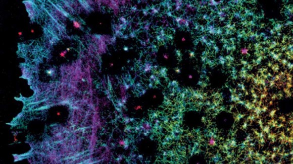
Popularization of super-resolution imaging techniques has allowed cell biologists to probe cell structure and function in previously unattainable detail. These methodologies continue to evolve, with new improvements that allow tailoring the available techniques to a particular need and application. This collection showcases primary research articles, reviews and protocols and highlights these recent developments by exemplifying the new, interesting applications of super-resolution microscopy as well as related tool development. We hope that this compilation of works will inspire future research with the aim to resolve outstanding challenges and further expand the utility of super-resolution imaging across biological and medical disciplines.
This Perspective reviews nanoscopy via stimulated emission depletion (STED), focusing on challenges for biologists and how technical advances are helping to meet these challenges.
An axon's function is dictated by its morphology and, thus, by the properties and organization of the axonal cytoskeleton. Leterrieret al. describe how advances in super-resolution and live-cell imaging are transforming our understanding of the molecular architecture of the axonal shaft.
Fluorescence nanoscopy enables the optical imaging of cellular components with resolutions at the nanometre scale. With the growing availability of super-resolution microscopes, nanoscopy methods are being increasingly applied. Quantitative, multicolour, live-cell nanoscopy and the corresponding labelling strategies are under continuous development.
Synaptic vesicles participate in neuronal communication by storing and releasing neurotransmitter molecules. The neurotransmitters can be detected using electrochemistry and mass spectrometry, and vesicle structural elements can be detected by super-resolution microscopy. This Review describes these analytical techniques and how they unravel the mechanisms of cell communication.
In the last decade, super-resolution microscopy has enabled us to image cells with unprecedented detail. This Perspective discusses how the field can move beyond images to extract quantitative biological information from multidimensional SMLM data.
In this Review, De Nizet al. discuss the contribution of key imaging tools to advances in our understanding of Plasmodiumspp. biology and host–pathogen interactions over the past decade. These advances, pertaining to parasite structure and motility, as well as the liver and blood stages, have led to paradigm shifts in our knowledge of malaria.
Optical and force nanoscopy enable visualization of intracellular and extracellular microbial structures with unprecedented resolution. This Review discusses the principles, advantages and limitations of the main optical and force nanoscopy techniques available.
Formation of inter-organelle contacts between mitochondria and lysosomes, regulated by lysosomal RAB7 GTP hydrolysis, allows for bidirectional regulation of mitochondrial and lysosomal dynamics.
The authors show that Munc13-1 molecules form multiple supramolecular self-assemblies that serve as vesicular release sites. Having multiple Munc13-1 assemblies affords a stable synaptic weight, which confers robustness of synaptic computation.
In budding yeast, glucose withdrawal, via the Rag GTPases, leads to TORC1 inhibition through its re-organization into a giant, vacuole-associated helix named a TOROID (TORC1 organized in inhibited domain).
Shi et al. map the ciliary transition zone by STORM imaging, characterizing protein arrangements in nested rings and finding that mutations in RPGRIP1L that are associated with the ciliopathy Joubert syndrome disrupt SMO ciliary localization.
Salmonella Typhimurium E3 ligase LUBAC generates linear polyubiquitin patches in the ubiquitin coat that serves as a signalling platform for the recruitment of Optineurin and Nemo for xenophagy and local activation of NF-κB, respectively.
Deubiquitinase OTULIN targets linear (M1-linked) ubiquitin chain patches on cytosolic Salmonella Typhimurium to modulate NEMO, IKKα/IKKβ and NF-ĸB signalling and regulate secretion of pro-inflammatory cytokines and bacterial proliferation.
Using large-scale, super-resolution microscopy, Sochacki et al. define the spatial organization of 19 proteins within clathrin-coated pits during distinct stages of clathrin-mediated endocytosis.
Using structured illumination microscopy, Beach et al. and Hu et al. visualize the assembly of myosin II filaments in cells, describing a filament-partitioning mechanism, and long-range self-organization of filaments, respectively.
Bertocchi and colleagues describe the organization of cadherin-based adhesions using super-resolution microscopy. They find that α-catenin is important for vinculin localization and observe a conformational change in vinculin following its activation.
A spatial resolution of 30 nm (=λ/31) exceeding the diffraction limit is achieved by super-resolution fluorescence microscopy. The nanoscopic imaging scheme can be applied to coherent quantum-mechanical systems such as quantum dots, as well as colour centres.
The ability to quantify the organization of cell membrane molecules is limited by the density of labeling and experimental conditions. Here, the authors use super-resolution optical fluctuation (SOFI) for molecular density and clustering analyses, and investigate nanoscale distribution of CD4 glycoprotein.
Studying interactions between lysosomes and mitochondria in living cells is difficult due to the limitations of existing probes. Here, the authors develop new cell-permeable fluorescent probes to image the dynamics of lysosomes and their physical interactions with mitochondria using super-resolution microscopy.
An automated system for data acquisition and analysis enables high-content screening localization microscopy and increases the throughput and information content of super-resolution microscopy methods such as dSTORM, DNA-PAINT and (spt)PALM.
Upconversion nanoparticles, which do not suffer from the photophysical artifacts that limit fluorescent molecules, offer an exciting opportunity for biological super-resolution imaging. Here, Zhan et al. develop an efficient STED mechanism using optimized lanthanide upconversion nanoparticles, enabling cytoskeleton nanoscopic imaging.
Ratiometric fluorescent pH probes are useful tools to monitor acidification of vesicles during endocytosis, but the size of vesicles is below the diffraction limit. Here the authors develop a family of ratiometric pH sensors for use in STED super-resolution microscopy, and optimize their delivery to endosomes.
Expansion microscopy, a technique for super-resolution imaging, is extended to clinical human tissue samples that are formalin fixed, paraffin embedded, stained and/or fresh frozen.
The measurement of molecular diffusion at sub-diffraction scales has been achieved in 2D space using STED-FCS, but an implementation for 3D diffusion is lacking. Here the authors present an analytical approach to probe diffusion in 3D space using STED-FCS and measure the diffusion of EGFP at different spatial scales.
In DNA-PAINT, transient binding of dye-labeled oligonucleotides to their target strands creates the ‘blinking’ required for stochastic nanoscopy. This protocol describes how to apply DNA-PAINT, from sample preparation to data processing.
Iterative expansion microscopy (iExM) is a strategy that achieves high resolution expansion microscopy by expanding samples multiple times. Expanding a sample twice enables ∼4.5 × 4.5 ∼20× physical expansion and ∼25 nm resolution.
This protocol describes the preparation of calibration bead slides, their use and additional strategies to reduce artifacts of structured illumination microscopy that will allow researchers to exploit the technique's full potential for biological applications.
This protocol describes how to prepare samples for labeling nuclei of cultured mammalian cells for 3D structured illumination microscopy of nuclear structures. Image acquisition, registration and downstream image analysis are also described.
Four different techniques for preparing and acquiring super-resolution CLEM data sets on aldehyde-fixed specimens are provided: Tokuyasu cryosectioning; whole-cell mount; cell unroofing and platinum replication; and resin embedding and sectioning.
New fluorescent biosensors enable the first super-resolution imaging of enzyme activity in live cells via fluorescence fluctuation increase by contact (FLINC).
Classical physics enabled subdiffraction-limited imaging has rarely been extended to the quantum regime. Here, Israelet al. develop a super-resolution localization microscopy based on non-classical photon statistics, enabling optical tracking of multiple quantum emitters.
Super-resolution optical microscopy based on stimulated emission depletion effects can now be performed at much lower light intensities than before by using bright upconversion emission from thulium-doped nanoparticles.
Stimulated emission double depletion addresses the issue of background in super-resolution imaging and quantitative microscopy through implementation of a two-pulse sequence in a modified stimulated emission depletion set-up. The measured background intensity is removed from each voxel in the acquired images thanks to time-resolved detection.
This protocol describes a detailed method for superresolution imaging of plant tissues by structured illumination microscopy (SIM). Details include microscope calibration, tissue preparation, image acquisition and evaluation of SIM images.
Spectacular images of the process of myosin II filament formation and organization in migrating cells are unveiled by super-resolution imaging. A combination of short- and long-range interactions with actin filaments is seen to play a critical role in filament partitioning and alignment into contractile actin arcs and stress fibres.
The ability to switch fluorophores on and off is key to performing super-resolution nanoscopy. To date, all switching schemes have been based on an incoherent response to the laser field. Now, a nanoscope that uses on–off coherent switching of quantum dots has been demonstrated.
Cadherin adhesion complexes have recently emerged as sensors of tissue tension that regulate key developmental processes. Super-resolution microscopy experiments now unravel the spatial organization of the interface between cadherins and the actin cytoskeleton and reveal how vinculin, a central component in cadherin mechanotransduction, is regulated by mechanical and biochemical signals.