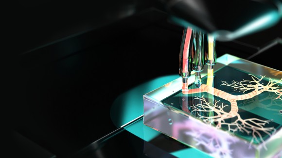
Thank you for visiting nature.com. You are using a browser version with limited support for CSS. To obtain
the best experience, we recommend you use a more up to date browser (or turn off compatibility mode in
Internet Explorer). In the meantime, to ensure continued support, we are displaying the site without styles
and JavaScript.
Modelling human tissues in microphysiologically relevant ‘chips’ will increasingly help to unravel mechanistic knowledge underlying disease, and might eventually accelerate the productivity of drug development and predict how individual patients will respond to specific drugs.
Multi-chambered vascularized human cardiac organoids produced under anisotropic stress and integrated with sensors facilitate the study of electro-metabolic coupling in arrythmia.
A human intestine-on-a-chip lined by intestinal epithelium derived from patients with environmental enteric dysfunction and cultured in nutrient-deficient medium recapitulates essential features of intestinal dysfunction associated with the disease.
The metabolically driven maturation of geometrically aligned human cardiac microtissues on a microfabricated chip reduces cell-phenotype-dependent variabilities in the action-potential morphology and calcium handling of the cardiomyocytes.
Tissue chips with matured human heart, liver, bone and skin tissue niches linked by recirculating vascular flow recapitulate interdependent functions of these organs.
This Perspective highlights notable milestones from the past two decades of research in cardiac tissue engineering, and outlines opportunities and challenges for further advancement.
Tissues and organoids derived from human pluripotent stem cells with knocked-out kinesin-2 subunits lack cilia, and can be used to model ciliopathy phenotypes and to reveal underlying mechanisms of disease.
A protocol for the production of human spinal-cord-like organoids that recapitulate the tube-forming morphogenesis of the early human spinal cord facilitates screening for antiepileptic drugs that can cause neural-tube defects.
Patterned organoids and bioprinted tissues can be generated by simultaneously co-differentiating pluripotent stem cells into distinct cell types via the forced overexpression of transcription factors, independently of culture-media composition.
Organoid models of intestinal stem cell differentiation into Paneth cells allow for the identification, via high-throughput phenotypic screening, of biological targets and small molecules regulating the composition of intestinal epithelium.
A microphysiological model of ischaemic stroke consisting of a functional neurovascular unit on a microfluidic chip allows for the systematic characterization of the neurorestorative outcomes of candidate stem cell therapies.
A microwell chip facilitates the single-cell characterization of the differentiation of aggregates of human induced pluripotent stem cells into pancreatic duct-like organoids and the discovery of secreted markers of pancreatic carcinogenesis.
A neurovascular-unit-on-a-chip with a functional blood–brain barrier recapitulates the neurotropism and barrier penetration of the most common pathogen causing fungal meningitis.
A microfluidic bronchial-airway-on-a-chip lined by human bronchial-airway epithelium and pulmonary endothelium can be used to rapidly identify antiviral therapeutics and prophylactics with repurposing potential.
Thousands of individual gastrointestinal organoids cultured on microcavity arrays without a solid extracellular matrix allow for high-throughput drug screening and for high-content image-based phenotypic analyses.
Cardiac organoids incorporating an oxygen-diffusion gradient and stimulated with the neurotransmitter noradrenaline can model the structure and function of the human heart after myocardial infarction.
This Review summarizes progress in the development of engineering strategies employed in reproductive science and medicine, with a focus on biomaterials and microfluidic approaches.
A vascularized human bone-marrow-on-a-chip improves the maintenance of patient-derived CD34+ cells, and recapitulates clinically relevant aspects of bone marrow injury as well as key haematopoietic defects of patients with a rare genetic disorder.
Physiological modelling of first-pass metabolism using data from a robotic instrument that fluidically links relevant organ chips predicts human pharmacokinetic parameters for orally administered nicotine and for intravenously injected cisplatin.
A system employing liquid-handling robotics and an integrated mobile microscope enables the automated culture, sample collection and in situ microscopy imaging of up to ten fluidically coupled organ chips within a standard tissue-culture incubator.
A microphysiological cartilage on a chip that enables the application of strain-controlled compression recapitulates the mechanical factors involved in the pathogenesis of osteoarthritis.
A microfluidic intestine-on-a-chip that allows the control of physiologically relevant oxygen gradients, enables the extended coculture of living human intestinal epithelium with stable communities of aerobic and anaerobic human gut microbiota.
A tumour-on-a-chip model featuring patient-derived glioblastoma cells, vascular endothelial cells and decellularized extracellular matrix from brain tissue can be used to identify patient-specific resistance to standard chemoradiotherapy.
A microphysiological model of the bronchial airways enables the study of the mechanochemical feedback interactions between smooth muscle cells and epithelial cells that underlie bronchospasm.
An endothelialized microfluidic system that recapitulates physiological properties of the microvasculature enables the real-time visualization of vascular-pathology features associated with sickle-cell disease and malaria, with high spatiotemporal resolution.
An efficient and chemically defined protocol for the differentiation of human induced pluripotent stem cells into podocytes enables the recapitulation of the differential clearance of the human kidney glomerulus in an organ-on-a-chip.
Pharmaceutical companies continue to advocate for the use of in vitro models towards the reduction of animal use in drug discovery and development while acknowledging that further advancements are needed to heighten the models’ current state of readiness.
Better cell sourcing and increasingly fine control over cell differentiation, tissue formation and cell and tissue maturation are pushing forward progress in disease modelling, drug development and regenerative medicine.
A robotic culture system for the high-throughput analysis of drug transport in porcine gastrointestinal tissue explants accurately predicts the absorption of orally taken drugs in the human gut.
Structural and functional changes following infarction in a human heart can be modelled with human cardiac organoids set in a hypoxic gradient and stimulated with the neurotransmitter noradrenaline.
Modelling human tissues in microphysiologically relevant ‘chips’ will increasingly help to unravel mechanistic knowledge underlying disease, and might eventually accelerate the productivity of drug development and predict how individual patients will respond to specific drugs.
An osteoarthritis model in a cartilage-on-a-chip, enabled by hyperphysiological compression, recapitulates the progression of the disease and its response to drugs.
A microfluidic chip incorporating oxygen gradients, a diverse human microbiota and patient-derived cells, mimics interactions between microorganisms and host tissue in the human gut.
A bioprinted glioblastoma-on-a-chip model enables the evaluation of treatment responses for individual patients whose brain tumours resist standard chemoradiotherapy.
A physiologically relevant microvasculature-on-a-chip device enables the study of microvascular pathology associated with inflammation and haematological diseases.
The efficient generation of mature podocytes from induced pluripotent stem cells makes possible the recapitulation of renal blood filtration on a chip.