Thank you for visiting nature.com. You are using a browser version with limited support for CSS. To obtain
the best experience, we recommend you use a more up to date browser (or turn off compatibility mode in
Internet Explorer). In the meantime, to ensure continued support, we are displaying the site without styles
and JavaScript.
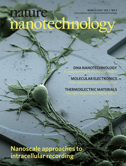
Action potentials have a central role in the nervous system. Intracellular methods can record these potentials with high signal-to-noise ratios, but they are invasive, whereas extracellular methods suffer from reduced signal strength. In this issue three groups report advances in this field. Lieber and co-workers show that nanowire field-effect transistors can make electrical measurements on biological materials with unprecedented spatial resolution. Cui and colleagues use arrays of vertical nanopillar electrodes to make both intra- and extracellular recordings with excellent signal strength and minimal damage to the cells. And Park and co-workers show that arrays of vertical silicon nanowires can record and stimulate neuronal activity from within mammalian nerve cells, such as the rat cortical cell in this false-colour SEM image, and also study the connections between these cells. The nanowire array is below the cell and cannot be seen in this image (which is 45 μm across).
Two independent groups have demonstrated that nanoscale electrodes can record action potentials in neurons and cardiac muscle cells, and a third group has shown that nanowire field-effect transistors can make electrical measurements on biological materials with unprecedented spatial resolution.
The spin-dependent Peltier effect has been demonstrated in a nanostructure consisting of a non-magnetic metal sandwiched between two ferromagnetic layers.
Using two gold nanoparticles to connect an antibody to metal electrodes results in the formation of a molecular junction that is both stable and highly reproducible.
A nanowire attached to an optical fibre can deliver payloads or light into specific compartments within a living cell, and also detect optical signals from subcellular regions with high spatial resolution.
A single point defect in graphene can act as an atomic antenna in the petahertz frequency range, leading to surface plasmon resonances at the subnanometre scale.
A silicon nanowire field-effect transistor coupled to the interior of a cell by means of a hollow silicon dioxide nanotube can detect changes in the electric potential of the intracellular fluid.
Arrays of vertical silicon nanowires can record and stimulate neuronal activity from within mammalian nerve cells, and can also map multiple individual synaptic connections between these cells.
Arrays of vertical nanopillar electrodes can be used for both intracellular and extracellular recording with excellent signal strength and quality, and minimal damage to the cells.
A nanowire waveguide attached to an optical fibre can deliver payloads into cells and act as an endoscope capable of imaging single living cells with high spatial resolution.
A single molecule of the antibody immunoglobulin G can self-assemble with two gold nanoparticles to fabricate a protein transistor in a highly reproducible manner.