Volume 7
-
No. 12 December 2012
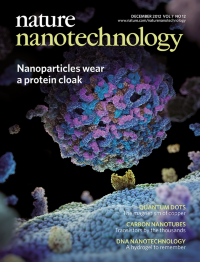
Unless specifically designed to avoid it, nanoparticles adsorb biomolecules from their surroundings when they enter the biological environment. This corona of biomolecules forms the identity of the nanoparticles because it is this surface that interacts with biological systems. The importance of this corona has been seen in biomaterial implants and cell scaffolds. However, unlike macroscopic surfaces, nanoparticles can travel to almost every part of the organism and further exchange biomolecules along its path. In the Review by Kenneth Dawson and co-workers, they discuss the basic concepts of this corona and how its properties may be linked to the biological behaviour of nanoparticles. Key issues are outlined for future research. The concept is illustrated on the cover that shows a corona on a nanoparticle surface interacting with a receptor protruding from the cell lipid bilayer.
Article p779
IMAGE: M.MONOPOLI, C. ÅBERG AND THE SCIENCE PICTURE COMPANY
COVER DESIGN: ALEX WING
-
No. 11 November 2012
Cancer initiation and progression are characterized by complex molecular and structural changes in cells and the extracellular matrix. These changes are expected to affect the mechanical properties and responses of cells and their surrounding environment. The atomic force microscope (AFM) has been used to show that cancer cells are more compliant than healthy cells. However, the relevance of these single-cell measurements performed on cells isolated from tumours has been questioned because they lack the appropriate 3D tissue environment. Marija Plodinec et al. have now used an indentation-type AFM for the nanomechanical characterization and diagnosis of breast cancer progression. This is illustrated on the cover image that shows an AFM tip probing an invasive cancer cell that is protruding above a malignant matrix-embedded cluster.
Article p757; News & Views p691
IMAGE: EVA BIELER, MARIJA PLODINEC, RODERICK LIM; ARTWORK: MARTIN OEGGERLI/MICRONAUT 2012
COVER DESIGN: ALEX WING
-
No. 10 October 2012
The electrocatalytic properties of metal nanoparticles are typically probed by measuring the total electrocatalytic reaction current of a large number of particles. However, the catalytic activity of a nanoparticle can vary depending on its precise size, shape and composition, and therefore techniques are required that can rapidly interrogate individual nanoparticles. Shan and co-workers have now shown that a plasmonic-based electrochemical current imaging technique can simultaneously image and quantify the electrocatalytic reactions of individual nanoparticles. This electrochemical current image, which measures approximately 1μm across, is of a single platinum nanoparticle with a diameter of 80nm. Protons are reduced to dihydrogen at the surface of the nanoparticle, which causes changes in the local refractive index of the solution and allows the local current density to be monitored from the surface plasmon resonance signal.
Letter p668; News & Views p615
IMAGE: XIAONAN SHAN ET AL.
COVER DESIGN: ALEX WING
-
No. 9 September 2012
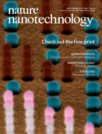
The ultimate resolution for printing colour images is dictated by the diffraction limit of visible light. To achieve this limit, Joel K. W. Yang and co-workers from IMRE, A*STAR in Singapore use a nanopatterned surface composed of silverâgold nanoposts and a backreflector over a silicon substrate. The metallic nanostructures interact with the incident light of a bright-field microscope through surface plasmon resonances and reflect a wide range of colours depending on the diameter and spacing of the nanoposts. The cover shows a false colour close-up of this nanopatterned surface coding for a portion of the left eye in the famous Lena image. The colours of the nanoposts correspond to the actual reflected wavelength, as seen through a bright-field microscope.
Letters p557; News & Views p550
IMAGE: KARTHIK KUMAR, HUIGAO DUAN, RAVI S. HEGDE, SAMUEL C. W. KOH, JENNIFER N. WEI AND JOEL K. W. YANG
COVER DESIGN: ALEX WING
-
No. 8 August 2012
A magnetic shift register consists of magnetic domains that can be moved in a controlled fashion, and can be used for logic and memory devices. Nanoscale variants are particularly promising, but have required very high current densities, highly restricted pathways or complex rotating magnetic fields. Now researchers at Eindhoven University of Technology have made a magnetic shift register in which domains move along arbitrary pathways, including closed loops, and are driven by an alternating magnetic field fixed in one direction. The cover image shows six frames from a simulation of this shift register, with red and blue colours representing different magnetization directions. The domain boundaries in the z-direction (long bars) are seen to move continually from left to right.
Letters p499
IMAGE: J. H. FRANKEN
COVER DESIGN: ALEX WING
-
No. 7 July 2012
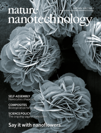
Nanoscale structures that are shaped like flowers have been routinely produced from inorganic materials for almost a decade. Now Jun Ge, Jiandu Lei and Richard Zare have made nanoflowers from a combination of an inorganic material (copper phosphate) and various organic materials (in the form of proteins). The increased surface area offered by their flower-like structure endows these hybrid nanoflowers with many useful properties. In particular, using an enzyme as the protein can lead to enhanced activity and stability, making such enzyme nanoflowers suitable for applications in catalysis and biosensing. This scanning electron micrograph, which measures 28 μm across, shows nanoflowers in which the organic material is laccase.
Letters p428; News & Views p415
IMAGE: JUN GE
COVER DESIGN: ALEX WING
-
No. 6 June 2012
Nanoparticles can deliver a variety of cancer drugs directly into tumour cells, which increases the efficacy of the treatment while reducing side effects. Small interfering RNAs (siRNAs) have shown promise as therapeutic agents but it is difficult to get them into cells. Now Daniel Anderson and co-workers have demonstrated that self-assembled DNA nanoparticles can reliably deliver siRNAs into cells and silence target genes in tumours. As shown in this illustration, the DNA nanoparticles are tetrahedral in shape, with six protruding arms. DNA nanoparticles have a number of properties that are useful for drug-delivery applications: it is relatively easy to control their size and to bind either drug molecules or targeting ligands to them.
Letters p389; News & Views p344
IMAGE: HYUKJIN LEE
COVER DESIGN: ALEX WING
-
No. 5 May 2012
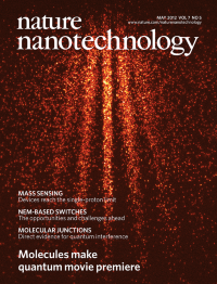
The observation of interference patterns in double-slit experiments with massive particles is generally regarded as the ultimate demonstration of the quantum nature of these particles. Moreover, unlike what happens in classical physics, it is possible to watch the build up of the pattern as the particles arrive at the detector one by one. Now researchers from Vienna, Tel Aviv and Basel have used a combination of nanofabrication and nanoimaging to record these patterns building up in real time for molecules with a mass of 1,298â AMU, which is a new record for such experiments, and 514â AMU (see cover; image measures 320â μm across). Last year many of the same researchers observed quantum interference of even heavier molecules (6,910â AMU) but did not record the build up of the interference pattern in thoseâ experiments.
Letters p297; News & Views p277
IMAGE: UNIV. OF VIENNA
COVER DESIGN: ALEX WING
-
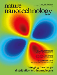
No. 4 April 2012
The ability to study the distribution of electric charge inside molecules would be useful in many areas of science and technology. Now researchers at IBM Research-Zurich have used a technique called Kelvin probe force microscopy to image the charge distribution inside a single naphthalocyanine molecule on a surface. This molecule has two hydrogen atoms at its centre and four lobes that give it a cross shape. The IBM team shows that the two lobes parallel to the hydrogen atoms have a lower charge density than the other two lobes. This computer simulation shows the asymmetry in the electric field above the molecule and perpendicular to the surface: regions of high field are shown in red and yellow; regions of low field are shown in blue. The image measures just over 2 nm across.
Letters p227; News & Views p210
IMAGE: FABIAN MOHN, IBM RESEARCH-ZURICH
COVER DESIGN: ALEX WING
-
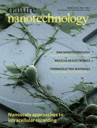
No. 3 March 2012
Action potentials have a central role in the nervous system. Intracellular methods can record these potentials with high signal-to-noise ratios, but they are invasive, whereas extracellular methods suffer from reduced signal strength. In this issue three groups report advances in this field. Lieber and co-workers show that nanowire field-effect transistors can make electrical measurements on biological materials with unprecedented spatial resolution. Cui and colleagues use arrays of vertical nanopillar electrodes to make both intra- and extracellular recordings with excellent signal strength and minimal damage to the cells. And Park and co-workers show that arrays of vertical silicon nanowires can record and stimulate neuronal activity from within mammalian nerve cells, such as the rat cortical cell in this false-colour SEM image, and also study the connections between these cells. The nanowire array is below the cell and cannot be seen in this image (which is 45 μm across).
Letters p174, p180 and p185; News & Views p143
IMAGE: J. T. ROBINSON, M. JORGOLLI, F. FRANKEL AND H. PARK
COVER DESIGN: ALEX WING
-
No. 2 February 2012
Nanopore-based sensors are being developed for possible applications in DNA sequencing. These sensors normally work by recording the ionic current through the nanopore: in particular they record the small changes in the current caused by the passage of biomolecules through the nanopore. However, DNA molecules pass through nanopores extremely quickly and the bandwidth of existing read-out systems is not high enough to cope with the resulting signal. Now Charles Lieber and co-workers have combined solid-state nanopores with nanowire field-effect transistors to create a high bandwidth read-out system in which the presence of a DNA molecule changes the electric potential near the nanopore, which in turn modulates the conductivity of the nanowire. The small rectangle at the centre of this photograph is a silicon nitride membrane (about 90 μm across) that contains the nanowire-nanopore sensors. The white lines are metal electrodes.
Letter p119; News & Views p81
IMAGE: PING XIE, HARVARD UNIVERSITY
COVER DESIGN: ALEX WING
-
No. 1 January 2012
DNA-based assembly can be used to create ordered three-dimensional arrays of inorganic nanoparticles in which the nanoparticles are held in place by short strands of DNA that are attached to their surface. Using two different types of nanoparticles allows a wider variety of binary superlattices to be created. Now Chad Mirkin and co-workers have shown that replacing some of the nanoparticles with hollow spacer particles made only of DNA allows an even wider variety of structures to be created, including one that has never been seen before. This artist's impression shows a hexagonal superlattice formed by replacing some of the gold nanoparticles (red spheres; not to scale) in an AB2 lattice with spacer nanoparticles (grey spheres). The background image is a small-angle X-ray diffraction pattern.
Letter p24
IMAGE: JOSHUA I. CUTLER, KE ZHANG, AND EVELYN AUYEUNG
COVER DESIGN: ALEX WING