
Volume 28 Issue 6, June 2023
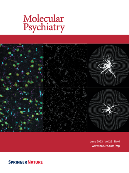
Immunofluorescent detection of astrocytes (Gfap; magenta) and neurons (NeuN; yellow) with DAPI (nuclei mark) in the basolateral amygdala of adult rats exposed during adolescence to vehicle (top panels) or high-dose THC (bottom panels). Middle images show skeletonized Gfap processes, and right panels are representative astrocyte sholl images. For more information see the article by Ferland et al. on pages 2583–2593.
Image
-
Advertisement
