Figures, tables and video
From the following article
Surgical therapy for gastroesophageal reflux disease
Renee C. Minjarez and Blair A. Jobe
GI Motility online (2006)
doi:10.1038/gimo56
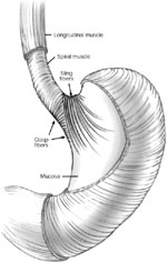
Figure 1
Opposing sling and clasp muscle fibers. The longitudinal muscle layer of the stomach has been cut away to show the opposing sling and clasp muscle fibers.
Full size figure and legend (63K)
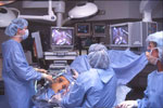
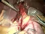
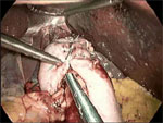
Figure 3
A typical operating room setup for performing laparoscopic antireflux surgery (LARS).
Full size figure and legend (124K)
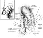
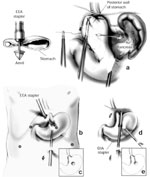
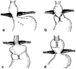

Table 1
Laparoscopic versus open fundoplication results of four randomized controlled trials
Full size table and legendTable 2
Laparoscopic versus open Nissen fundoplication results of four randomized controlled trials
Full size table and legendTable 4
Results of laparoscopic Nissen fundoplication in 181 patients followed for 5 years
Full size table and legend