
Figure 4 - Nissen fundoplication.
From the following article
Surgical therapy for gastroesophageal reflux disease
Renee C. Minjarez and Blair A. Jobe
GI Motility online (2006)
doi:10.1038/gimo56
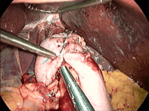
This intraoperative view is looking superiorly toward the hiatus and shows a complete Nissen fundoplication wrapping posteriorly around the distal esophagus. The grasper to the left (patient's right) points to three sutures holding the two ends of the fundus securely over the distal esophagus. The liver is seen being retracted superiorly by a laparoscopic liver retractor, an essential piece of equipment for exposing the hiatus. The grasper in the middle shows the course of the esophagus as it enters the abdomen. The crura are not seen in this picture. (Source: Courtesy of B.A. Jobe, M.D.)
Powerpoint slides for teaching
If the slide opens in your browser, Select "File > Save as" to save it.
Download Power Point slide (1,980K)