Thank you for visiting nature.com. You are using a browser version with limited support for CSS. To obtain
the best experience, we recommend you use a more up to date browser (or turn off compatibility mode in
Internet Explorer). In the meantime, to ensure continued support, we are displaying the site without styles
and JavaScript.
Method of the Year 2023: methods for modeling development
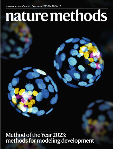
Methods for modeling development are our pick for the Method of the Year 2023. The cover shows mouse blastocysts stained for trophectoderm (cyan), epiblast (yellow) and primitive endoderm (magenta).
Research with human embryos and embryo models, this year’s Method of the Year, can be fraught. In contrast, digital embryos could be studied, even perturbed, in computational what-happens-when experiments.
The creation of multiple stem-cell-derived models of mammalian embryogenesis is opening many new doors to study human development and brings a need for scientists to demonstrate responsible dialog over the associated ethical issues.
In vivo developmental atlases provide a crucial reference for the new class of stem-cell-derived human embryo models, helping accelerate insights into the mechanisms of human development.
Advancements in methods that enable in vitro culture of mammalian embryos have become an essential way of investigating mammalian early embryonic development and modeling developmental and pregnancy-related disorders. Here, we discuss the recent method development in this space and analyze current challenges and future directions.
Recent methodological advances in measurements of geometry and forces in the early embryo and its models are enabling a deeper understanding of the complex interplay of genetics, mechanics and geometry during development.
Increasingly advanced in vitro stem-cell-derived human embryo models raise novel ethical questions and shed a light on long-standing questions regarding research on human embryos.
DARLIN enables the generation of a massive diversity of barcodes for in vivo lineage tracing and the combination with single-cell multi-omics measurements.
A class of protein-based molecular shape probes move us closer toward the goal of a general, genetically encoded tagging system for cryogenic electron tomography.
Fluorescent actinometers enable the measurement of light intensity even in the depths of samples and over wide ranges of wavelengths and intensities. We introduce two protocols to quantitatively characterize the spatial distribution of light of various fluorescence imaging systems and to calibrate the illumination of commercially available instruments and light sources.
Cardinal v.3 is an open-source software for reproducible analysis of mass spectrometry imaging experiments, and includes data processing features such as mass recalibration, statistical analyses such as single-ion segmentation and rough annotation-based classification, and analyses of large-scale multitissue experiments.
An engineered RNA A-to-I deaminase (rABE) offers low sequence bias, high activity and low background for REMORA (RNA-encoded molecular recording in adenosines) and enables improved molecular recording of RNA–protein interactions.
Genetically encoded multimeric particles (GEMs) are 25-nm tags with recognizable structural signatures, which can be used to label specific proteins in mammalian cells to facilitate their subcellular localization in cryo-ET.
nextPYP is a turn-key framework for single-particle cryo-electron tomography that streamlines complex data analysis pipelines, from pre-processing of tilt series to high-resolution refinement, for efficient analysis and visualization of large datasets.
Adaptable, turn-on maturation (ATOM) biosensors use monobody or nanobody targeting to control fluorescent protein maturation for fluorescence in the presence of target biomolecules, enabling bright and specific cellular biosensing.
Two methods for fluorescence-based actinometry using organic dyes and photoconvertible fluorescent proteins enable rapid and precise measurement of light intensity at the sample in fluorescence microscopes.
DBlink uses deep learning to capture long-term dependencies between different frames in single-molecule localization microscopy data, yielding super spatiotemporal resolution videos of fast dynamic processes in living cells.
Enhanced super-resolution radial fluctuations (eSRRF) offers improved image fidelity and resolution compared to the popular SRRF method and further enables volumetric live-cell super-resolution imaging at high speeds.
DeepSeMi is a self-supervised denoising framework that can enhance SNR over 12 dB across diverse samples and imaging modalities. DeepSeMi enables extended longitudinal imaging of subcellular dynamics with high spatiotemporal resolution.
A pulsed illumination scheme renders stimulated Brillouin microscopy less phototoxic and allows imaging of the mechanical properties of sensitive samples such as single cells, Caenorhabditis elegans embryos, zebrafish larvae and organoids.
SegCLR automatically annotates segmented electron microscopy datasets of the brain with information such as cellular subcompartments and cell types, using a self-supervised contrastive learning approach.