
Thank you for visiting nature.com. You are using a browser version with limited support for CSS. To obtain the best experience, we recommend you use a more up to date browser (or turn off compatibility mode in Internet Explorer). In the meantime, to ensure continued support, we are displaying the site without styles and JavaScript.

Volume 2 Issue 12, December 2005

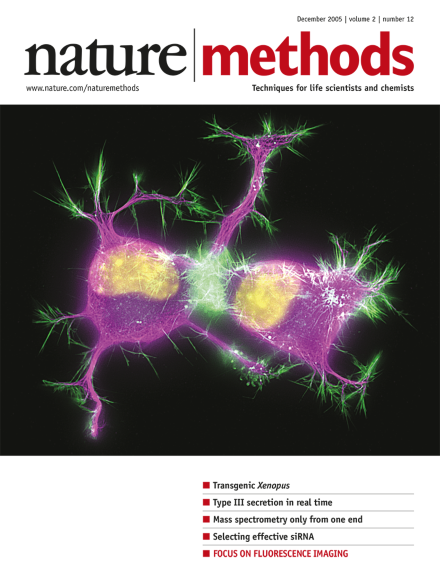
Cytoskeleton in differentiating neuroblastoma cells. Image courtesy of Torsten Wittmann, Department of Cell and Tissue Biology, University of California, San Francisco.

Editorial

Top of page ⤴

Research Highlights

Top of page ⤴

News & Views

  • A Xenopus embryo coinjected with a plasmid encoding a transgene and the φC31 integrase mRNA readily facilitates genomic integration resulting in healthy transgenic embryos.

    • Stuart J Smith
    • Timothy J Mohun
    News & Views
  • The pathogenic arsenal of many bacteria includes an apparatus that mediates the injection of a cocktail of virulence proteins directly into host cells. Spatiotemporal aspects of this process can now be analyzed in living cells.

    • Luís Jaime Mota
    • David W Holden
    News & Views
Top of page ⤴

Foreword

Top of page ⤴

Commentary

  • Fluorescence microscopy has undergone a renaissance in the last decade. The introduction of green fluorescent protein (GFP) and two-photon microscopy has allowed systematic imaging studies of protein localization in living cells and of the structure and function of living tissues. The impact of these and other new imaging methods in biophysics, neuroscience, and developmental and cell biology has been remarkable. Further advances in fluorophore design, molecular biological tools and nonlinear and hyper-resolution microscopies are poised to profoundly transform many fields of biological research.

    • Rafael Yuste
    Commentary
Top of page ⤴

Perspective

Top of page ⤴

Review Article

Top of page ⤴

Brief Communication

Top of page ⤴

Article

Top of page ⤴

Protocol

Top of page ⤴

Technology Feature

  • Human genotyping has never been hotter, and a sophisticated set of array-based tools now simplifies the process dramatically, facilitating everything from small basic research studies to complex genetic epidemiology. Alan Dove reports.

    • Alan Dove
    Technology Feature
Top of page ⤴

Classic Protocol

  • Northern blotting and hybridization are used to study gene expression by detecting RNA species of interest, and to identify alternate RNA splicing patterns. It is analogous to Southern blotting, which is used to analyze DNA. This protocol describes the transfer of RNA from agarose gels to nylon membranes and the fixation of the RNA to the membrane1,2,3.

    Classic Protocol
Top of page ⤴

Advertising Feature: Application Note

Top of page ⤴

Focus

  • A series of review articles and other content discussing the principles of fluorescence and different imaging modalities along with practical advice.

    Focus
Top of page ⤴

Search

Quick links