Volume 19
-
No. 12 December 2022
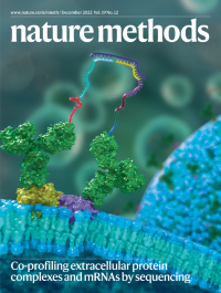
Co-profiling extracellular protein complexes and mRNAs by sequencingProximity-sequencing (Prox-seq) uses DNA-barcoded antibody probes to detect proteins and their pairwise complexes on the surface of single cells.
See Vistain et al.
-
No. 11 November 2022
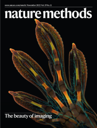
The beauty of imagingThe winning image of the Nikon Small World 2022 Photomicrography Competition, an embryonic foot of a Madagascar giant day gecko (Phelsuma grandis). The image was captured using whole-mount fluorescence staining, tissue clearing, high-resolution confocal microscopy and image stitching.
See Editorial
-
No. 10 October 2022
Focus on methods for studying noncoding RNAThis month we present a Focus on methods for studying noncoding RNA and future directions for deciphering the regulatory roles of noncoding RNA. The confetti conceptually illustrates the broad diversity of noncoding RNA and the complexity of their biological implications.
See Editorial
-
No. 9 September 2022
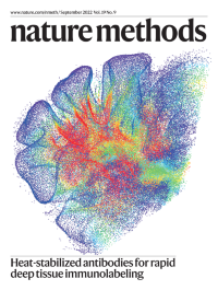
Heat-stabilized antibodies for rapid deep tissue immunolabelingHeat-stabilized antibodies (SPEARS) enable thermally facilitated 3D immunolabeling (THiCK staining) of parvalbumin-expressing cells in a mouse cerebellar hemisphere.
See Lai et al.
-
No. 8 August 2022
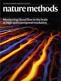
Monitoring blood flow in the brain at high spatiotemporal resolutionFunctional ultrasound localization microscopy reveals whole-brain vascular changes during neuronal activation at high resolution, providing quantitative information on changes in flow, speed and vessel diameter in multiple vascular compartments over a wide field of view.
See Renaudin et al.
-
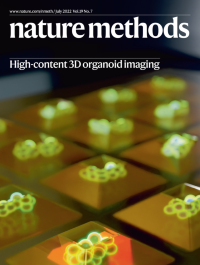
No. 7 July 2022
High-content 3D organoid imagingJeWell microchips facilitate compartmentalized organoid culture and allow single-objective light sheet imaging of up to 96 organoids in 3D and in three colors in one hour.
See Beghin et al.
-
No. 6 June 2022
Tools for assembling and analyzing complete genomesWith new tools developed by the Telomere-to-Telomere (T2T) Consortium, the human genome is revealed in greater quality and detail.
See Editorial
-
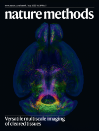
No. 5 May 2022
Versatile multiscale imaging of cleared tissuesOn the cover, an optically cleared mouse brain imaged with a hybrid open-top light-sheet microscope.
See Glaser et al.
-
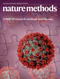
No. 4 April 2022
COVID-19 research: methods lead the wayDecades of accumulated methods development across diverse areas of basic biological research have facilitated a speedy scientific response to the SARS-CoV-2 virus.
See Editorial
-
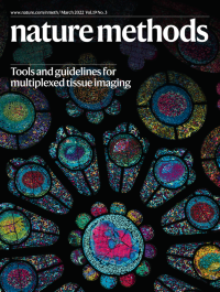
No. 3 March 2022
Tools and guidelines for multiplexed tissue imagingIBEX (iterative bleaching extends multiplexity) imaging of cell–cell interactions in a human lymph node evokes a stained glass window in a cathedral.
See Hickey et al.
-
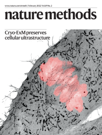
No. 2 February 2022
Cryo-ExM preserves cellular ultrastructureA human cell in mitosis observed using cryo-expansion microscopy (Cryo-ExM). The DNA is stained pink and the rest of the cell with an NHS ester that marks the proteome and highlights the mitochondria in black at each pole of the mitotic spindle.
See Laporte et al.
-
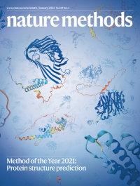
No. 1 January 2022
Method of the Year 2021: Protein structure predictionProtein structure prediction is our Method of the Year 2021, for the recent development of deep-learning-based methods that predict structures with unprecedented accuracy. On the cover, a blizzard of protein structure models from the AlphaFold Protein Structure Database (https://alphafold.ebi.ac.uk/), predicted by the method AlphaFold2.
See Editorial