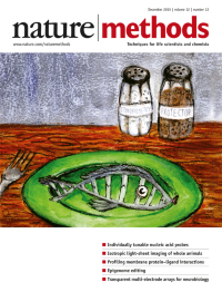
Volume 12
-
No. 12 December 2015
A double-stranded DNA probe with its 'flavor' adjusted by stoichiometry. The relative concentration of a protector oligonucleotide controllably modulates the probe's specificity and sensitivity. Cover by Cindy Thaung. Article p1191
-
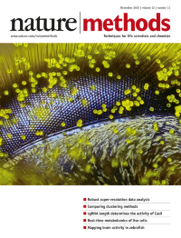
No. 11 November 2015
Eye of a honeybee covered in dandelion pollen (120x magnification), photographed using reflected light illumination and constructed from about 150 separate stacked images by Ralph Claus Grimm, Jimboomba, Queensland, Australia. Winner of the 2015 Nikon Small World photomicrography contest (reprinted with permission of R.C.G.; image provided by Nikon).
-
No. 10 October 2015
A mouse depicted on a radio-frequency transducer. The transducer activates optogenetic responses in the animal through an implanted light-delivery system. Cover by Erin Dewalt. Article p969
-
No. 9 September 2015
The MIME (mutational interference mapping experiment) method maps sequence and secondary-structure motifs critical for RNA function. Cover by Erin Dewalt, based on a figure from Smyth et al. Article p866
-
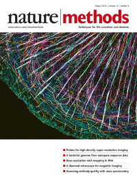
No. 8 August 2015
IRIS probes yield high-density labeling for high-fidelity super-resolved imaging of cytoskeletal components. Cover by Tai Kiuchi, Kyoto University Faculty of Medicine. Brief Communication p743
-
No. 7 July 2015
A human-liver chimeric mouse infected with human malaria parasites replaces chimpanzees as a host for conducting experimental genetic crosses. Original artwork by Asa Pefferman, Center for Infectious Disease Research. Brief Communication p631
-
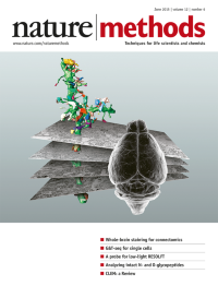
No. 6 June 2015
Reconstruction of a spiny dendrite and incident synaptic boutons from serial block-face electron microscopy data taken from a brain prepared with the BROPA method. Cover by Shawn Mikula (Max Planck Institute for Neurobiology) and Julia Kuhl (http://somedonkey.com/). Article p541
-
No. 5 May 2015
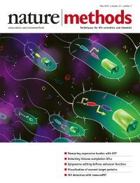
GFP output can be used to measure the burden that heterologous gene expression exerts on engineered Escherichia coli cells. Cover by Yutong Wu, Imperial College London. Brief Communication p415
-
No. 4 April 2015
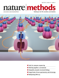
Commercial nanopore sequencing works by detecting changes in electrical current as individual long DNA molecules transit a protein nanopore with the help of an accessory enzyme. Cover by Erin Dewalt. Article p351.
-
No. 3 March 2015
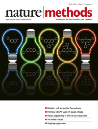
Chemical structures of bright organic fluorophores containing a novel azetidine group. The colors of the lightbulb outlines match the emission maxima of the fluorophores.Original artwork by Luke Lavis, Howard Hughes Medical Institute/Janelia Farm Research Campus. Cover by Erin Dewalt. Article p244.
-
No. 2 February 2015
Extracellular matrix patterned in a mesh consisting of 20-μm-wide stripes of laminin (green) and fibronectin (purple) spaced 20 μm apart and overlaid conformally on highaspect ratio microridges. Image by Q. Jallerat and J.M. Szymanski, Carnegie Mellon University. Article p134.
-
No. 1 January 2015
Light-sheet fluorescence microscopy is our choice for Method of the Year 2014 for its ability to image three dimensional biological samples at high speed and with low toxicity. Cover design by Erin Dewalt. Light-sheet image from Nik962/iStock/Thinkstock. Special feature begins on p19.