Thank you for visiting nature.com. You are using a browser version with limited support for CSS. To obtain
the best experience, we recommend you use a more up to date browser (or turn off compatibility mode in
Internet Explorer). In the meantime, to ensure continued support, we are displaying the site without styles
and JavaScript.
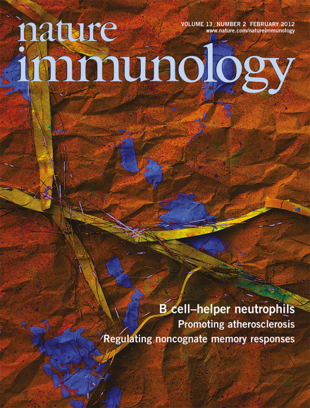
Under homeostatic conditions, B cell–helper neutrophils colonize the perifollicular area of the spleen to stimulate antibody diversification and production in marginal zone B cells, as reported by Puga and colleagues (p 170; and News & Views by Tangye & Brink, p 111). The original confocal microscopy image shows splenic B cell–helper neutrophils forming neutrophil extracellular trap–like B cell–interacting projections that express the pattern-recognition receptor CEACAM1 (green) and the glycoprotein CD15 (red). DAPI (blue) stains DNA. Original image by Linda Cassis. Artwork by Lewis Long.
A workshop organized by the Society for Leukocyte Biology offered advice to young scientists on how to decipher the grant-submission process of the US National Institutes of Health and compose a clear, compelling and fundable grant.
Since the definition of an immunoselective proteasome, the 'immunoproteasome', its function has been heavily disputed. A mouse with complete deficiency in immunoproteasomes has resolved one issue of the adaptation of the immune system to the proteasome but has raised other questions.
Neutrophils and B cells are thought to specialize in driving innate and adaptive immune responses to infection. New data suggest that these cells may also communicate in the splenic marginal zone to drive T cell–independent antibody responses.
IFN-γ is a cytokine that is critical for defense against intracellular bacterial pathogens. New work provides evidence that the recognition of bacterial flagellin by the NLRC4 inflammasome in splenic dendritic cells triggers rapid release of IL-18, which leads to IFN-γ production by memory CD8+ T cells.
AhR is more than just a receptor for pollutants. Studies have now identified crucial roles for this environment-sensing transcription factor in the development and maintenance of gut-associated lymphoid tissues.
Immunoproteasomes incorporate three alternative β subunits that catalyze peptide cleavage. Rock and colleagues generate mice lacking all three immunoproteasome-specific subunits and reveal marked differences in MHC class I presentation and CD8+ T cell responses.
Lipid-engorged macrophages accumulate in atherosclerotic plaques where chronic inflammation ensues. Moore and colleagues show that macrophage migration to chemokines and egress is arrested by netrin-1, which is secreted in the plaque lesions.
The generation of certain gut innate lymphoid cell (ILC) populations requires the aryl hydrocarbon receptor (AHR). Colonna and colleagues show that the induction of Notch expression by AHR is required for the development of interleukin 22–producing NKp46+ ILCs.
How signals derived from dendritic cells regulate T cell lineage fate remains unclear. Chi and colleagues identify p38α in dendritic cells as a central integrator of instructive signals for TH17 differentiation.
Noncognate CD8+ memory T cells can be rapidly activated by bacteria. Bedoui and colleagues show that this process is specifically dependent on recognition by and activation of the NLRC4 inflammasome.
Follicular T cells provide help to B cells to elicit antibody responses. Cerutti and colleagues show that neutrophils provide help to marginal-zone B cells that produce T cell–independent antibodies.
Interferon-α is a critical mediator of pathogen-induced thymic involution. Liston and colleagues show that the microRNA miR-29a reduces sensitivity of thymic epithelium to infection signals and protects against thymus involution.
Chronically infected mice upregulate expression of inhibitory molecules on exhausted T cells. Harty and colleagues report similar findings in human patients with malaria and show that blockade of the inhibitory receptors PD-L1 and LAG-3 restores antimalaria responses in mice.