Thank you for visiting nature.com. You are using a browser version with limited support for CSS. To obtain
the best experience, we recommend you use a more up to date browser (or turn off compatibility mode in
Internet Explorer). In the meantime, to ensure continued support, we are displaying the site without styles
and JavaScript.
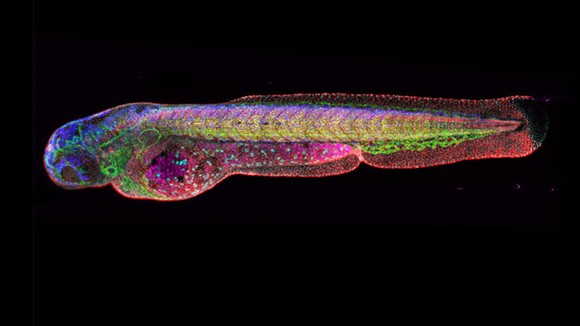
One of the goals of biological imaging is to watch biological processes where they occur – within living tissue or even within a living animal. But in vivo imaging presents a set of challenges that are not encountered when imaging relatively small, flat samples like cells. In this Focus issue, we bring together papers on methods for optical imaging within living tissue. Two Reviews discuss: microscopy methods for functional brain imaging and the principles and practicalities of the different flavours of light sheet microscopy. A Perspective describes the use of adaptive optics to correct aberrations in scattering samples. This issue also includes primary research papers on multiphoton microscopy, an editorial that reminds our readers of the many areas of this large and exciting field that we do not cover, and a collection of recent papers on deep imaging from Nature Research journals.
Yang and Yuste review currently available technologies for optical imaging of neural circuits, comparing them to help researchers choose optimal ones for their applications.
This Review introduces the fundamental considerations for building a light sheet microscope, describes the pros and cons associated with available implementations, and offers practical advice for users.
Ouzounov et al. report calcium imaging with three-photon microscopy in the mouse brain. The approach enabled noninvasive recording of activity with high spatial and temporal resolution from GCaMP6-labeled neurons located as deep as the hippocampus.
vTwINS enables high-speed volumetric calcium imaging via a V-shaped point spread function and a dedicated data-processing algorithm. Song et al. apply this strategy to image population activity in the mouse visual cortex and hippocampus.