
Thank you for visiting nature.com. You are using a browser version with limited support for CSS. To obtain the best experience, we recommend you use a more up to date browser (or turn off compatibility mode in Internet Explorer). In the meantime, to ensure continued support, we are displaying the site without styles and JavaScript.

Volume 15 Issue 6, June 2020

Cryo-FIB milling for structural biology in cells

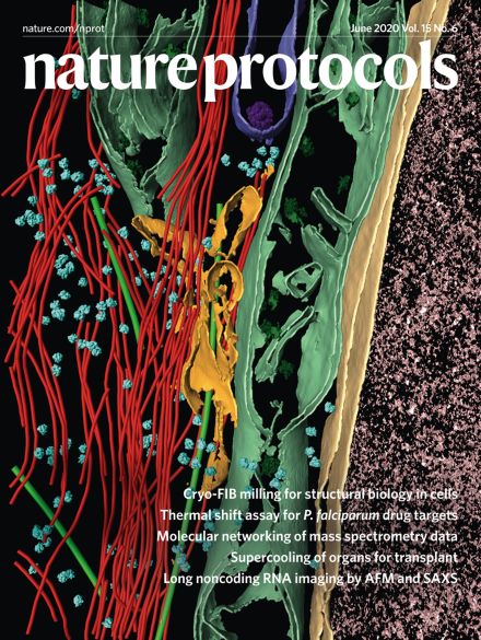
3-D segmentation of a cryo-electron tomogram of a cryo-FIB milled human U2OS cell. Various organelles and cytoskeletal elements are shown, e.g., ribosomes in cyan, actin and intermediate filaments in red, microtubules in dark green, mitochondria in light green, nuclear envelope in yellow and nucleoplasm in pink.

See Wagner et al.

Image: Reika Watanabe and Robert Buschauer. Cover Design: Erin Dewalt

Editorial

  • Nature Protocols is pleased to be a part of the Resource Identification Initiative, a project aimed at improving the reproducibility of research by clearly identifying key biological resources. Stable unique digital identifiers, called Research Resource Identifiers (RRIDs), are assigned to individual resources, allowing users to accurately identify and source them, track their history, identify known problems (such as cell line contamination and misidentification) and find relevant research papers. Following a successful 6-month trial, we will now require authors to provide RRIDs for all antibodies and cell lines used in their protocols. We will also be encouraging them to add RRIDs for other tools (such as plasmids and organisms) where they think this is helpful.

    Editorial

    Advertisement

Top of page ⤴

Protocols

Top of page ⤴

Amendments & Corrections

Top of page ⤴

Search

Quick links