Thank you for visiting nature.com. You are using a browser version with limited support for CSS. To obtain
the best experience, we recommend you use a more up to date browser (or turn off compatibility mode in
Internet Explorer). In the meantime, to ensure continued support, we are displaying the site without styles
and JavaScript.
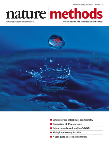
Releasing intact membrane protein complexes in bicelles or nanodiscs into the gas phase for observation by mass spectrometry. Photograph and cover art by Jonathan Hopper, Karl Harrison and Michelle Smikle. Brief Communication p1206
GFP targeted to satellite repeats via transcription activator–like effectors (TALEs) allows the imaging of nuclear dynamics early in mouse development.
Imaging with better than 200-nanometer resolution brings new subcellular-scale details into focus. Practitioners share how they weigh trade-offs in speed, resolution and phototoxicity.
RNA-seq is a recent and immensely popular technology for cataloging and comparing gene expression. Two papers from the international RGASP consortium report on large-scale competitions to identify the best algorithms for RNA-seq analysis, with surprising variability in the results.
This Perspective describes statistical measures commonly used to quantify whether nodes in biological networks have similar interaction profiles and discusses which indices are best suited for specific tasks.
The RGASP consortium compared 25 RNA-seq analysis programs in their ability to identify exons, reconstruct transcripts and quantify expression levels. Assembly of isoforms and their expression levels in higher eukaryotes remains a challenge.
Authors compare RNA-seq aligners on mouse and human data sets using benchmarks such as alignment yield, splice junction accuracy and suitability for transcript reconstruction. The work highlights the strength of each program and discusses outstanding needs in RNA-seq analysis.
Discrepancies between model prediction and experimental measurements enable molecular-level discovery with a whole-cell model of Mycoplasma genitalium.
A method and software for profiling microbial communities from shotgun sequence data uses universal single-copy marker sequences for accurate species-level assignment. The method can classify species lacking a reference genome sequence, making it possible to analyze the large fraction of unknown microbes in the human gut.
The metagenomeSeq tool robustly detects the differential abundance of microbes in marker-based microbial surveys by tackling the problems of data sparsity and undersampling common to these data sets.
A simple method for preparing entirely monovalent quantum dots is described. These reagents can be targeted to protein and lipid tags and used as imaging probes in live cells.
Amphipols, bicelles and nanodiscs are used to study intact membrane protein complexes by mass spectrometry, with better preservation of oligomeric complexes than traditional detergent micelles.
A database of known drug-gene interactions, with information derived from many public sources, allows the identification of genes that are currently targeted by a drug and the membership of genes in a category, such as kinase genes, that have a high potential for drug development.
The Spinach2 RNA aptamer provides substantially improved imaging over its predecessor Spinach, enabling live-cell imaging of many tagged RNA species including toxic trinucleotide repeat–containing RNAs.
This paper describes a platform for high-throughput image-based profiling of human pluripotent stem cells (hPSCs). It is used to quantify differentiation bias and endogenous signaling levels in hPSC lines.
Changes to protein interactomes as a result of mutations to the bait protein or addition of a pharmacological inhibitor are robustly monitored with affinity purification coupled with data-independent acquisition–based mass spectrometry and an automated data analysis pipeline. Also in this issue, Collins et al. describe a similar method.
Dynamic changes to the 14-3-3 protein interactome are robustly followed over time using affinity-purification data-independent analysis–based mass spectrometry. Also in this issue, Lambert et al. describe a similar method.