Volume 3
-
No. 12 December 2007
Using single-molecule fluorescence resonance energy transfer (smFRET), Kim et al. (p 763) demonstrate that the 8-17 DNAzyme, one of the newest members of the metalloenzyme family, uses both induced fit and lock-and-key modes of catalysis, much like its proteinaceous relatives (see also News and Views by Schlosser and Li, p 753). Shown is an smFRET image beneath a schematic representation of DNAzyme catalysis, with the 8-17 DNAzyme strand in blue and the substrate in red(labeled with a FRET donor in yellow and acceptor in orange). The path on the left demonstrates global folding that occurs prior to cleavage and release of the product. The path on the right depicts binding to a prearranged DNA structure. Cover art by Erin Boyle, based on imagery provided by Alex D. Jerez, Hee-Kyung Kim and Yi Lu.
-
No. 11 November 2007
A new nitric oxide signaling messenger. Sawa et al. (p 727) have demonstrated the cellular formation of 8-nitroguanosine 3',5'-cyclic monophosphate (8-nitro-cGMP), a 'nitrated' second messenger that is induced by nitric oxide (NO). 8-NitrocGMP not only behaves as a mimic for cyclic GMP (cGMP) and activates standard NO signaling pathways, but it also produces a unique protein sulfhydryl modification in which adduction of cGMP moieties to cysteine residues in proteins (called S-guanylation) takes place. This study now sheds light on an as-yet unrecognized area of NO second messenger and oxidative stress pathways (see also News & Views by Feelisch, p 687). The cover shows an immunofluorescence image of HepG2 cells that were treated with an NO donor (SNAP) and visualized for 8-nitro-cGMP (red) and S-guanylation (green; merged image in orange). Cover art by Erin Boyle, based on an image provided by Tatsuya Okamoto and Takaaki Akaike.
-
No. 10 October 2007
Being able to make time- and space-keeping measurements such as when to stop dividing and when to die is essential for both cells and whole organisms. This issue features articles that examine how chemical and biological systems make measurements and regulate their own space and time. Revealing how organisms regulate events spatially and temporally brings our understanding of the cell and its individual processes to a more mechanistic level. Cover art by Erin Boyle, based on photographs by Andrzej Pastuszak and Mike Wade.
Focus
-
No. 9 September 2007
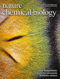
Natural product biosynthesis in vitro. Cheng et al. (p 557) report the in vitro total synthesis of the bacteriostatic natural product enterocin from Streptomyces maritimus. Balibar et al. (p 584) report the full in vitro reconstitution of the bisindole alkaloid terrequinone A, a fungal cytotoxic metabolite. Both papers highlight the power of assembling biosynthetic pathways outside of the cell (see also News & Views by Fecik, p 531). A picture of Aspergillus nidulans, the source of terrequinone A, is shown. Cover art by Erin Boyle, based on an image provided by Paul D. Straight and Carl J. Balibar.
-
No. 8 August 2007
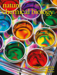
Besides its importance in drug discovery, high-throughput screening is increasingly being used in academic research to identify chemical probes of biological processes. In this issue, we feature a collection of articles that highlight the current state of screening. Cover art by Erin Boyle, based on a photograph from Getty Images.
-
No. 7 July 2007
Natural products, and terpenes in particular, have long fascinated scientists with their remarkable structural diversity and their often unknown biological functions. In this issue, we feature a collection of articles meant to shed light on the synthesis, sources and significance of terpenoid natural products. Terpene structures courtesy of Seiichi Matsuda. Cover art by Erin Boyle, based on a photo by Rodolfo Clix.
Focus
-
No. 6 June 2007
Dynamic O-GlcNAc glycosylation in the brain. Khidekel et al. (p. 339) have developed a method for determining levels of O-GlcNAc modification using quantitative mass spectrometry-based proteomics. The authors found that O-GlcNAc levels at specific protein sites are modulated in neurons and in rat brains in response to stimulation (see also News & Views by Wells, p 303). The cover shows the structure of the translation initiation factor eIF4G, which was found to be glycosylated in neurons, with a chemical model of the O-GlcNAc modification. In the background is an MRI image of a brain. The eIF4G structure is from http://www.pdb.org (PDB ID 1UG3), and the MRI brain image is from the Corbis Corporation (© Allen Bell/Corbis). Cover art by Erin Boyle, based on images provided by Linda Hsieh-Wilson and Peter M. Clark.
-
No. 5 May 2007
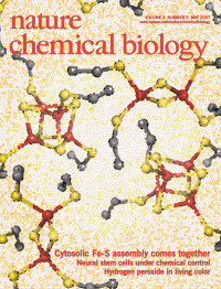
Iron-sulfur cluster assembly in the eukaryotic cytosol. Netz et al. (p 278) have identified the Cfd1-Nbp35 complex as a scaffold protein for iron-sulfur cluster assembly in the yeast cytosol. The Cfd1-Nbp35 complex can bind up to three [4Fe-4S] clusters and transfer them, both in vitro and in vivo, to target [Fe-S] apoproteins (see also News & Views by Broderick, p 243). Images of [2Fe-2S] and [4Fe- 4S] clusters are shown. The [2Fe-2S] image is from the [2Fe-2S] ferredoxin from Spirulina platensis (PDB ID 4FXC) and the [4Fe-4S] image is from Azotobacter vinelandii ferredoxin (PDB ID 1FDA); both images were prepared using PyMOL. Cover art by Erin Boyle, based on images provided by David Mulder, John Peters and Joan Broderick.
-
No. 4 April 2007
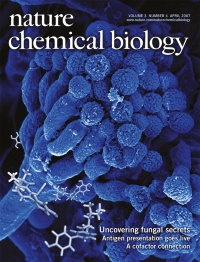
Activating silent gene clusters. Bergmann et al. have characterized metabolites encoded by a silent gene cluster in the Aspergillus nidulans genome. Expression of a pathway-specific activator induced the expression of a cryptic hybrid polyketide-nonribosomal synthase gene cluster and led to the discovery of the novel pyridone-containing metabolites aspyridone A and aspyridone B. An image of A. nidulans is shown, along with the structures of aspyridones A and B. Cover art by Erin Boyle, based on images and structures provided by Axel Brakhage and Christian Hertweck.
-
No. 3 March 2007
A clickable inhibitor reveals context-dependent autoactivation of p90 RSK. Cohen et al. (p 156) generated fmk-pa, a potent inhibitor of the p90 ribosomal S6 kinases RSK1 and RSK2. The authors used click chemistry to show specific and irreversible modification of RSKs in mammalian cells. RSKs are activated by the MEK-ERK signal transduction pathway through phosphorylation of the C-terminal kinase domain (CTD), which goes on to phosphorylate and activate the N-terminal kinase domain (NTD). Using their inhibitor, the authors found evidence for an unidentified kinase that bypasses RSK CTD activity. The cover illustrates CTD inhibition by fmk-pa, which should have cellular effects that are distinct from those of NTD inhibition by the recently reported compound BI-D1870 (see also News & Views by Frödin, p 138). Cover art by Erin Boyle, based on images and design provided by Michael Cohen.
-
No. 2 February 2007
Buey et al. (p 117) found that the natural product and microtubule stabilizing agent cyclostreptin binds covalently to microtubules first at an exposed site on the microtubule surface and then, upon passing through microtubule pores, at the paclitaxel binding site on the less accessible inner wall. Cyclostreptin effectively arrests the cell cycle in a multidrug-resistant tumor cell line. With the widespread use of microtubule stabilizing agents in cancer chemotherapy, such an irreversible agent has the potential to overcome the resistance characteristic of conventional antimitotic treatments because it can escape ejection from the cell by P-glycoprotein efflux pumps (see also News & Views by Snyder, p 81). Cover art by Erin Boyle, based on images provided by Isabel Barasoain, Oriol Pineda and Fernando Díaz.
-
No. 1 January 2007
RNA has occupied a central position in the central dogma of molecular biology, which holds that information flows from DNA through RNA to proteins. In this issue we feature a series of articles that focus on the rapidly moving field of RNA chemical biology. This month's cover highlights the diversity of RNA structure and function. RNA structural images, clockwise from the lower left: (1) sensing domain of the Escherichia coli thiamine pyrophosphate riboswitch (Serganov, A. et al. Nature 441, 1167-1171 (2006); PDB ID 2GDI); (2) phenylalanine tRNA from Saccharomyces cerevisiae (Westhof, E. & Sundaralingam, M. Biochemistry 25, 4868-4878 (1986); PDB ID 1TRA); (3) RNA silencing suppressor protein (p19) bound to a synthetic siRNA duplex (Vargason, J.M. et al. Cell 115, 799-811 (2003); PDB ID 1RPU); (4) hammerhead ribozyme (Scott, W.G. et al. Cell 81, 991-1002 (1995); PDB ID 1MME); (5) view of the RNA components of the large ribosomal subunit from Haloarcula marismortui (Ban, N. et al. Science 289, 905-920 (2000); PDB ID 1FFK). Image 1 courtesy of Alexander Serganov and Dinshaw Patel, Memorial Sloan-Kettering Cancer Center, New York, prepared using PyMOL and nuccyl. Images 2-5 courtesy of Jeffrey Vargason, George Fox University, Newberg, Oregon, USA, prepared using PyMOL. Cover art by Erin Boyle.
Focus