
Figures, tables and video
From the following article
Electromyography in oral and pharyngeal motor disorders
Adrienne L. Perlman
GI Motility online (2006)
doi:10.1038/gimo32
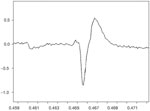
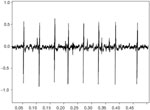
Figure 2
Train (MUAPT) of single MUAPs from the thyroarytenoid muscle displayed in Figure 1.
Full size figure and legend (31K)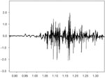
Figure 3
Firing pattern from a collection of MUAPs from the medial thyroarytenoid muscle.
Full size figure and legend (33K)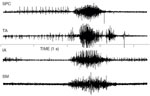
Figure 4
Simultaneous EMG recording from the superior pharyngeal constrictor (SPC), thyroarytenoid (TA), interarytenoid (IA), and submental muscles (SM) during swallow.
Full size figure and legend (46K)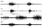
Figure 5
Simultaneous EMG recording of two swallows from the superior pharyngeal constrictor (SPC), thyroarytenoid (TA), cricopharyngeus (CP), and submental muscles (SM) during swallow.
Full size figure and legend (49K)
Figure 6
Rectified EMG recording from the superior pharyngeal constrictor muscles of a patient with unilateral pharyngeal paralysis.
Full size figure and legend (44K)