
Figures, tables and video
From the following article
Pathophysiology of achalasia and diffuse esophageal spasm
Ikuo Hirano
GI Motility online (2006)
doi:10.1038/gimo22
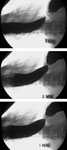
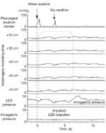
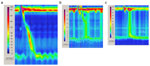
Figure 3
Contour plot topographic analysis of esophageal motility in achalasia.
Full size figure and legend (82K)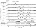
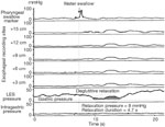
Figure 5
Esophageal manometric findings in achalasia variant with preserved LES relaxation.
Full size figure and legend (69K)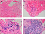
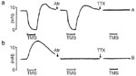
Figure 7
In vitro study demonstrating the effects of electrical field stimulation (EFS) on circular muscle strips from the LES of control subjects (a) and patients with achalasia (b).
Full size figure and legend (12K)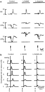
Figure 8
a: Effect of nitro-L-arginine methyl ester (L-NAME) on LES relaxation in the opossum in vivo.
Full size figure and legend (65K)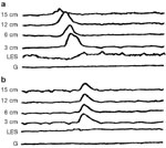
Figure 9
Effect of recombinant hemoglobin that inactivates nitric oxide on esophageal peristalsis in a human subject.
Full size figure and legend (54K)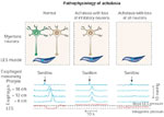
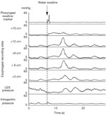
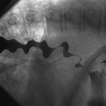
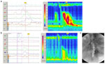
Figure 13
Contour plot topographic analysis of esophageal motility and esophagram in diffuse esophageal spasm.
Full size figure and legend (111K)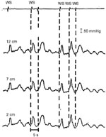
Figure 14
Failed deglutitive inhibition in diffuse esophageal spasm.
Full size figure and legend (56K)